

300
(Liu et al. 2002 ; Ju et al. 2003 ). Also, the new continuous cell line (KCF-1) from
caudal fi n of koi ( Cyprinus carpio koi ), that consists of short fi broblast-like cells,
was recently developed (Dong et al. 2012 ).
Unfortunately, relatively speaking only a few fi n cell lines were developed in
marine fi sh. For example, from fi n tissues of sheepshead ( Archosargus probatocephalus ) (Gregory et al. 1980 ), of gilt-head seabream ( Sparus aurata ) (Bejar
et al. 1997 ), of a grouper ( Epinephelus coioides ) (Chi et al. 1999 ), of a fl ounder
( Paralichthys olivaceus ) (Kang et al. 2003 ), of a tropical grouper ( Epinephelus awoara ) (Lai et al. 2003 ), from caudal fi n of brakish water fi sh Etroplus suratensis
(Swaminathan et al. 2010 ), from the a tail fi n (Imajoh et al. 2007 ) as well as from
dorsal fi n of red sea bream ( Pagrus major ) (Ku et al. 2010 ) have all been developed.
Of course, the idea to use fi sh fi n cell lines in tissue engineering with the
challenging task to develop corresponding fi sh-inspired robotic devices, seems to
be very speculative today. Indeed, it does beg the question: what is the state of the
art in fi sh robotics research today?
7.5 Robotic Fish-Like Devices
The diversity of form and design in fi shes is determined by tremendous evolutionary
success of these aquatic vertebrates (Fish 1992 ). Principally, the design specializations shown by fi sh are all still elaborations of features generic to the lower vertebrate classes (Katz 2002 ). During last decades, “biologists and engineers have made
considerable progress in understanding how animals like fi sh moving underwater use
their muscles to power movement. They have described body and appendage
motion during propulsion, and conduct experimental and computational analyses of
fl uid movement and attendant forces,” (Lauder et al. 2007 ).
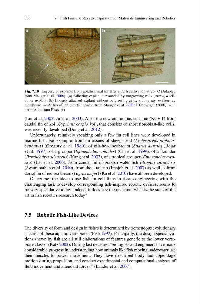
Fig. 7.10 Imagery of explants from goldfi sh anal fi n after a 72 h cultivation at 20 °C (Adapted
from Mauger et al. 2006 ). ( a ) Adhering explant surrounded by outgrowing cells ( arrow ) = celldonor explant. ( b ) Loosely attached explant without outgrowing cells. r bony ray, m inter-ray
membrane. Scale bar = 0.25 mm (Reprinted from Mauger et al. ( 2006 ), Copyright (2006), with
permission from Elsevier)
7 Fish Fins and Rays as Inspiration for Materials Engineering and Robotics
(Liu et al. 2002 ; Ju et al. 2003 ). Also, the new continuous cell line (KCF-1) from
caudal fi n of koi ( Cyprinus carpio koi ), that consists of short fi broblast-like cells,
was recently developed (Dong et al. 2012 ).
Unfortunately, relatively speaking only a few fi n cell lines were developed in
marine fi sh. For example, from fi n tissues of sheepshead ( Archosargus probatocephalus ) (Gregory et al. 1980 ), of gilt-head seabream ( Sparus aurata ) (Bejar
et al. 1997 ), of a grouper ( Epinephelus coioides ) (Chi et al. 1999 ), of a fl ounder
( Paralichthys olivaceus ) (Kang et al. 2003 ), of a tropical grouper ( Epinephelus awoara ) (Lai et al. 2003 ), from caudal fi n of brakish water fi sh Etroplus suratensis
(Swaminathan et al. 2010 ), from the a tail fi n (Imajoh et al. 2007 ) as well as from
dorsal fi n of red sea bream ( Pagrus major ) (Ku et al. 2010 ) have all been developed.
Of course, the idea to use fi sh fi n cell lines in tissue engineering with the
challenging task to develop corresponding fi sh-inspired robotic devices, seems to
be very speculative today. Indeed, it does beg the question: what is the state of the
art in fi sh robotics research today?
7.5 Robotic Fish-Like Devices
The diversity of form and design in fi shes is determined by tremendous evolutionary
success of these aquatic vertebrates (Fish 1992 ). Principally, the design specializations shown by fi sh are all still elaborations of features generic to the lower vertebrate classes (Katz 2002 ). During last decades, “biologists and engineers have made
considerable progress in understanding how animals like fi sh moving underwater use
their muscles to power movement. They have described body and appendage
motion during propulsion, and conduct experimental and computational analyses of
fl uid movement and attendant forces,” (Lauder et al. 2007 ).
Fig. 7.10 Imagery of explants from goldfi sh anal fi n after a 72 h cultivation at 20 °C (Adapted
from Mauger et al. 2006 ). ( a ) Adhering explant surrounded by outgrowing cells ( arrow ) = celldonor explant. ( b ) Loosely attached explant without outgrowing cells. r bony ray, m inter-ray
membrane. Scale bar = 0.25 mm (Reprinted from Mauger et al. ( 2006 ), Copyright (2006), with
permission from Elsevier)
7 Fish Fins and Rays as Inspiration for Materials Engineering and Robotics