

298
7.4 Fin Regeneration and Fin Cell Culture
Contrarily to mammals, adult teleost fi sh have the ability to regenerate their appendages
(Nakatani et al. 2007 ). Correspondingly, it is not surprising that studies on the
role of stem cells in tissue regeneration of fi sh are currently fashionable (see for
review Kawakami 2010 ).
As reviewed by Akimenko et al. ( 2003 ), a group of undifferentiated and proliferative
cells termed as blastema, is the key player in regeneration (see for details Santamaría
and Becerra 1991 ). A series of regulation mechanisms takes place between the cells
that are involved in the regeneration of corresponding part of the fi n. The simple
structure of the Teleosts fi n at the cellular level determines its fast regeneration.
Moreover, each fin ray exists as independent regeneration component. These
features make it an appropriate model to determine molecular mechanisms of
regeneration including different multiple pathways.
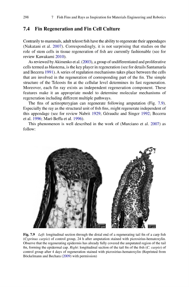
The fi ns of actinopterygian can regenerate following amputation (Fig. 7.9 ).
Especially the ray as the structural unit of fi sh fi ns, might regenerate independent of
this appendage (see for review Nabrit 1929 ; Géraudie and Singer 1992 ; Becerra
et al. 1996 ; Marí-Beffa et al. 1996 ).
This phenomenon is well described in the work of (Murciano et al. 2007 ) as
follow:
Fig. 7.9 Left : longitudinal section through the distal end of a regenerating tail fi n of a carp fi sh
( Cyprinus carpio ) of control group, 24 h after amputation stained with picrosirius-hematoxylin.
Observe that the regenerating epidermis has already fully covered the amputated region of the tail
fi n, forming the epidermal cap. Right : longitudinal section of the tail fi n of the fi sh ( C. carpio ) of
control group after 4 days of regeneration stained with picrosirius-hematoxylin (Reprinted from
Böckelmann and Bechara ( 2009 ) with permission)
7 Fish Fins and Rays as Inspiration for Materials Engineering and Robotics
7.4 Fin Regeneration and Fin Cell Culture
Contrarily to mammals, adult teleost fi sh have the ability to regenerate their appendages
(Nakatani et al. 2007 ). Correspondingly, it is not surprising that studies on the
role of stem cells in tissue regeneration of fi sh are currently fashionable (see for
review Kawakami 2010 ).
As reviewed by Akimenko et al. ( 2003 ), a group of undifferentiated and proliferative
cells termed as blastema, is the key player in regeneration (see for details Santamaría
and Becerra 1991 ). A series of regulation mechanisms takes place between the cells
that are involved in the regeneration of corresponding part of the fi n. The simple
structure of the Teleosts fi n at the cellular level determines its fast regeneration.
Moreover, each fin ray exists as independent regeneration component. These
features make it an appropriate model to determine molecular mechanisms of
regeneration including different multiple pathways.
The fi ns of actinopterygian can regenerate following amputation (Fig. 7.9 ).
Especially the ray as the structural unit of fi sh fi ns, might regenerate independent of
this appendage (see for review Nabrit 1929 ; Géraudie and Singer 1992 ; Becerra
et al. 1996 ; Marí-Beffa et al. 1996 ).
This phenomenon is well described in the work of (Murciano et al. 2007 ) as
follow:
Fig. 7.9 Left : longitudinal section through the distal end of a regenerating tail fi n of a carp fi sh
( Cyprinus carpio ) of control group, 24 h after amputation stained with picrosirius-hematoxylin.
Observe that the regenerating epidermis has already fully covered the amputated region of the tail
fi n, forming the epidermal cap. Right : longitudinal section of the tail fi n of the fi sh ( C. carpio ) of
control group after 4 days of regeneration stained with picrosirius-hematoxylin (Reprinted from
Böckelmann and Bechara ( 2009 ) with permission)
7 Fish Fins and Rays as Inspiration for Materials Engineering and Robotics