

293
biomineralize in a proximodistal direction within the extracellular collagenous network
of the basal lamella belonging to the epidermal-dermal interface of the fi n (Géraudie
and Landis 1982 ).
According to description of Geraudie and Landis ( 1982 ), “needle- and plate-like
particles of a solid mineral phase appear to be associated with the collagen fi brils
and with a fi ne, granular, interfi brillar material central to the demirays. Cellular
processes and membrane-bound vesicles are absent from the regions of calcifi cation. During fi n growth, the bony, acellular lepidotrichia are separated from the
epidermal-dermal interface by infi ltrating mesenchymal cells in proximal fi n
regions; in distal areas, the lepidotrichia remain within the basal lamella.
Considerations of embryologic and structural features of fi n components fail to
support the hypothesis that individual segments of lepidotrichia are modifi ed scales
in all fi sh,” (Géraudie and Landis 1982 ).
Recently, in experiments with developing zebrafi sh fi ns, it was shown that “the transcription factor Evx1 is expressed in the joints between individual lepidotrichia segments and at the distal tips of the lepidotrichia. It is also expressed in the apical growth
zone in regenerating fi ns. This is a very specifi c phenotype as both lepidotrichia hemisegment separations and lepidotrichia bifurcations still form normally in Evx1 mutant fi ns,
as do joints in the more proximal endoskeletal radials,” (Schulte et al. 2011 ).
Actinotrichia are located in brush-fashioned groups at the distal tip of each ray,
where they provide a fl exible support to the fi n edge being non-calcifi ed fusiform
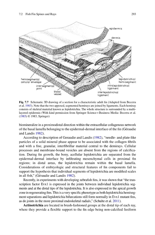
Fig. 7.7 Schematic 3D drawing of a section for a characteristic adult fi n (Adapted from Becerra
et al. 1983 ). Note that the two apposed, segmented hemirays are joined by ligaments. Each hemiray
consists of skeletal material known as lepidotrichia. The whole structure is surrounded by a multilayered epidermis (With kind permission from Springer Science + Business Media: Becerra et al.
( 1983 ) © 1983, Springer)
7.2 Fish Fin Spines and Rays
biomineralize in a proximodistal direction within the extracellular collagenous network
of the basal lamella belonging to the epidermal-dermal interface of the fi n (Géraudie
and Landis 1982 ).
According to description of Geraudie and Landis ( 1982 ), “needle- and plate-like
particles of a solid mineral phase appear to be associated with the collagen fi brils
and with a fi ne, granular, interfi brillar material central to the demirays. Cellular
processes and membrane-bound vesicles are absent from the regions of calcifi cation. During fi n growth, the bony, acellular lepidotrichia are separated from the
epidermal-dermal interface by infi ltrating mesenchymal cells in proximal fi n
regions; in distal areas, the lepidotrichia remain within the basal lamella.
Considerations of embryologic and structural features of fi n components fail to
support the hypothesis that individual segments of lepidotrichia are modifi ed scales
in all fi sh,” (Géraudie and Landis 1982 ).
Recently, in experiments with developing zebrafi sh fi ns, it was shown that “the transcription factor Evx1 is expressed in the joints between individual lepidotrichia segments and at the distal tips of the lepidotrichia. It is also expressed in the apical growth
zone in regenerating fi ns. This is a very specifi c phenotype as both lepidotrichia hemisegment separations and lepidotrichia bifurcations still form normally in Evx1 mutant fi ns,
as do joints in the more proximal endoskeletal radials,” (Schulte et al. 2011 ).
Actinotrichia are located in brush-fashioned groups at the distal tip of each ray,
where they provide a fl exible support to the fi n edge being non-calcifi ed fusiform
Fig. 7.7 Schematic 3D drawing of a section for a characteristic adult fi n (Adapted from Becerra
et al. 1983 ). Note that the two apposed, segmented hemirays are joined by ligaments. Each hemiray
consists of skeletal material known as lepidotrichia. The whole structure is surrounded by a multilayered epidermis (With kind permission from Springer Science + Business Media: Becerra et al.
( 1983 ) © 1983, Springer)
7.2 Fish Fin Spines and Rays