

282
and attach to the heads of the caudal fi n rays. There is no differentiation of superfi cial
and deep caudal musculature in Polypterus and no separation of distinct intrinsic
muscles,” (Lauder 1989 ).
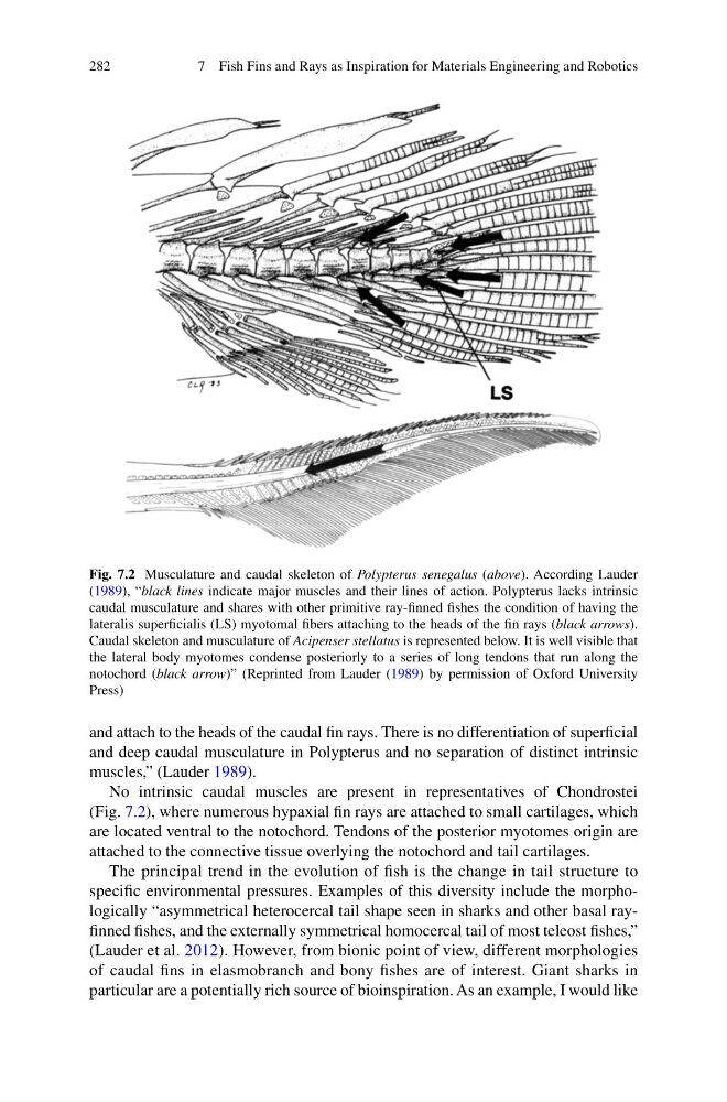
No intrinsic caudal muscles are present in representatives of Chondrostei
(Fig. 7.2 ), where numerous hypaxial fi n rays are attached to small cartilages, which
are located ventral to the notochord. Tendons of the posterior myotomes origin are
attached to the connective tissue overlying the notochord and tail cartilages.
The principal trend in the evolution of fi sh is the change in tail structure to
specifi c environmental pressures. Examples of this diversity include the morphologically “asymmetrical heterocercal tail shape seen in sharks and other basal rayfi nned fi shes, and the externally symmetrical homocercal tail of most teleost fi shes,”
(Lauder et al. 2012 ). However, from bionic point of view, different morphologies
of caudal fi ns in elasmobranch and bony fi shes are of interest. Giant sharks in
particular are a potentially rich source of bioinspiration. As an example, I would like
Fig. 7.2 Musculature and caudal skeleton of Polypterus senegalus ( above ). According Lauder
( 1989 ), “ black lines indicate major muscles and their lines of action. Polypterus lacks intrinsic
caudal musculature and shares with other primitive ray-fi nned fi shes the condition of having the
lateralis superfi cialis (LS) myotomal fi bers attaching to the heads of the fi n rays ( black arrows ).
Caudal skeleton and musculature of Acipenser stellatus is represented below. It is well visible that
the lateral body myotomes condense posteriorly to a series of long tendons that run along the
notochord ( black arrow )” (Reprinted from Lauder ( 1989 ) by permission of Oxford University
Press)
7 Fish Fins and Rays as Inspiration for Materials Engineering and Robotics
and attach to the heads of the caudal fi n rays. There is no differentiation of superfi cial
and deep caudal musculature in Polypterus and no separation of distinct intrinsic
muscles,” (Lauder 1989 ).
No intrinsic caudal muscles are present in representatives of Chondrostei
(Fig. 7.2 ), where numerous hypaxial fi n rays are attached to small cartilages, which
are located ventral to the notochord. Tendons of the posterior myotomes origin are
attached to the connective tissue overlying the notochord and tail cartilages.
The principal trend in the evolution of fi sh is the change in tail structure to
specifi c environmental pressures. Examples of this diversity include the morphologically “asymmetrical heterocercal tail shape seen in sharks and other basal rayfi nned fi shes, and the externally symmetrical homocercal tail of most teleost fi shes,”
(Lauder et al. 2012 ). However, from bionic point of view, different morphologies
of caudal fi ns in elasmobranch and bony fi shes are of interest. Giant sharks in
particular are a potentially rich source of bioinspiration. As an example, I would like
Fig. 7.2 Musculature and caudal skeleton of Polypterus senegalus ( above ). According Lauder
( 1989 ), “ black lines indicate major muscles and their lines of action. Polypterus lacks intrinsic
caudal musculature and shares with other primitive ray-fi nned fi shes the condition of having the
lateralis superfi cialis (LS) myotomal fi bers attaching to the heads of the fi n rays ( black arrows ).
Caudal skeleton and musculature of Acipenser stellatus is represented below. It is well visible that
the lateral body myotomes condense posteriorly to a series of long tendons that run along the
notochord ( black arrow )” (Reprinted from Lauder ( 1989 ) by permission of Oxford University
Press)
7 Fish Fins and Rays as Inspiration for Materials Engineering and Robotics