

171
3.6.3 Conclusion
A thorough overview on salt glands with attention on their nonhomologous origin has
been done by Nicolson and Lutz ( 1989 ). These supraorbitally located glands represent
one of the most effective organs in the vertebrate kingdom involved in the epithelial
transport of ions (sodium and chloride) against a marked concentration gradient
(Gerstberger and Gray 1993 ). Thus, the organs used as salt glands in different organisms are nonhomologous and have apparently evolved independently (Peaker and
Linzell 1975 ). Ion secretion occurs in the rectal glands of sharks (Burger and Hess
1960 ), the sublingual glands of snakes (Dunson 1968 ; Dunson et al. 1971 ), the nasal
glands of birds and lizards, and the postorbital glands of turtles (Peaker and Linzell
1975 ). Despite their nonhomologous origins, the salt glands of these different organisms are all characterized microscopically by principal cells with large quantities of
mitochondria and convoluted lateral, and sometimes basal, membranes (Ellis and Abel
1964 ; Peaker and Linzell 1975 ). The similarity in the structure of the salt glands of different species suggests that there may be similarities in salt gland operation as well.
There is evidence of hypertrophy in response to long- term increases in salt loading in
both birds and turtles (Peaker and Linzell 1975 ; Cowan 1969 , 1971 ). Unfortunately,
there is lack of knowledge about the visualization of biohalite production by secreting
cells at the nanolevel. However, I hope that the recently established method to obtain
primary culture of avian salt gland secretory cells (Lowy et al. 1989 ) could be used for
these kinds of experiments in the future. Furthermore, the biomineralogical community
needs data on both chemical composition and nanostructural organization of biohalite
that can be obtained from different taxa of marine vertebrates.
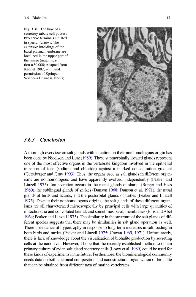
Fig. 3.31 The base of a
secretory tubule cell possess
two nerve terminals situated
in special furrows. The
extensive infoldings of the
basal plasma membrane are
localized in the upper part of
the image (magnifi cation × 50,000; Adapted from
Kühnel 1982, with kind
permission of Springer
Science + Business Media)
3.6 Biohalite
3.6.3 Conclusion
A thorough overview on salt glands with attention on their nonhomologous origin has
been done by Nicolson and Lutz ( 1989 ). These supraorbitally located glands represent
one of the most effective organs in the vertebrate kingdom involved in the epithelial
transport of ions (sodium and chloride) against a marked concentration gradient
(Gerstberger and Gray 1993 ). Thus, the organs used as salt glands in different organisms are nonhomologous and have apparently evolved independently (Peaker and
Linzell 1975 ). Ion secretion occurs in the rectal glands of sharks (Burger and Hess
1960 ), the sublingual glands of snakes (Dunson 1968 ; Dunson et al. 1971 ), the nasal
glands of birds and lizards, and the postorbital glands of turtles (Peaker and Linzell
1975 ). Despite their nonhomologous origins, the salt glands of these different organisms are all characterized microscopically by principal cells with large quantities of
mitochondria and convoluted lateral, and sometimes basal, membranes (Ellis and Abel
1964 ; Peaker and Linzell 1975 ). The similarity in the structure of the salt glands of different species suggests that there may be similarities in salt gland operation as well.
There is evidence of hypertrophy in response to long- term increases in salt loading in
both birds and turtles (Peaker and Linzell 1975 ; Cowan 1969 , 1971 ). Unfortunately,
there is lack of knowledge about the visualization of biohalite production by secreting
cells at the nanolevel. However, I hope that the recently established method to obtain
primary culture of avian salt gland secretory cells (Lowy et al. 1989 ) could be used for
these kinds of experiments in the future. Furthermore, the biomineralogical community
needs data on both chemical composition and nanostructural organization of biohalite
that can be obtained from different taxa of marine vertebrates.
Fig. 3.31 The base of a
secretory tubule cell possess
two nerve terminals situated
in special furrows. The
extensive infoldings of the
basal plasma membrane are
localized in the upper part of
the image (magnifi cation × 50,000; Adapted from
Kühnel 1982, with kind
permission of Springer
Science + Business Media)
3.6 Biohalite