

169
No other gland in higher vertebrates, except the mammalian kidney, can produce
fl uids which are concentrated to this degree. The concentration limit for electrolytes
in the kidney of man is about 400 mEq/L, in the rat 600 mEq/L, in the kangaroo
rat, 1,500 mEq/L, and in the champion concentrator, the North African rodent
Psammomys , 1,900 mEq/L (Schmidt-Nielsen 1960 ). Thus, the salt gland compares
favorably to the kidney in its concentrating ability.
Osmotically free water is the product generated due salt gland secretion. The phenomenon is briefl y reviewed by Hughes ( 2003 ) and Hughes et al. ( 2007 ) as follows:
“Acclimation to saline induces interstitial water and Na move into cells. When
the bird drinks seawater, Na enters the plasma from the gut, and plasma osmolality
(Osm(pl)) increases. This induces water to move out cells expanding the extracellular fl uid volume (ECFV). Both increases in Osm(pl) and ECFV stimulate salt
gland secretion. The augmented intracellular fl uid content should allow more rapid
expansion of ECFV in response to elevated Osm(pl), and facilitate activation of salt
gland secretion. To fully utilize the potential of the salt glands, intestinally absorbed
NaCl must be reabsorbed by the kidneys. Thus, Na uptake at gut and renal levels
may constrain extrarenal NaCl secretion,” (Hughes 2003 ).
3.6.2 Salt Glands: From Anatomy to Cellular Level
The common anatomical feature for all tetrapods is that their salt glands are localized
within cephalic area. However the anatomical position of these formations varies
among animal groups. Thus, according to Babonis and Brischoux ( 2012 ) three
following cephalic areas are “currently recognized:
(1) nasal glands in extinct archosaurs, extant birds, and lizards;
(2) orbital glands in turtles;
(3) oral glands in extant crocodiles and snakes,” (Babonis and Brischoux 2012 ).
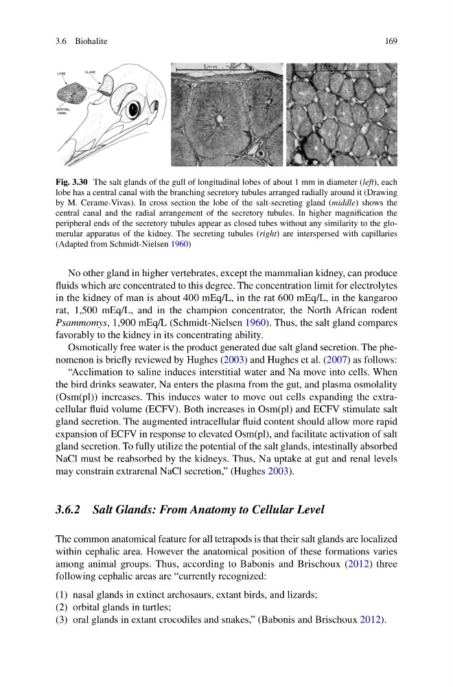
Fig. 3.30 The salt glands of the gull of longitudinal lobes of about 1 mm in diameter ( left ), each
lobe has a central canal with the branching secretory tubules arranged radially around it (Drawing
by M. Cerame-Vivas). In cross section the lobe of the salt-secreting gland ( middle ) shows the
central canal and the radial arrangement of the secretory tubules. In higher magnifi cation the
peripheral ends of the secretory tubules appear as closed tubes without any similarity to the glomerular apparatus of the kidney. The secreting tubules ( right ) are interspersed with capillaries
(Adapted from Schmidt-Nielsen 1960 )
3.6 Biohalite
No other gland in higher vertebrates, except the mammalian kidney, can produce
fl uids which are concentrated to this degree. The concentration limit for electrolytes
in the kidney of man is about 400 mEq/L, in the rat 600 mEq/L, in the kangaroo
rat, 1,500 mEq/L, and in the champion concentrator, the North African rodent
Psammomys , 1,900 mEq/L (Schmidt-Nielsen 1960 ). Thus, the salt gland compares
favorably to the kidney in its concentrating ability.
Osmotically free water is the product generated due salt gland secretion. The phenomenon is briefl y reviewed by Hughes ( 2003 ) and Hughes et al. ( 2007 ) as follows:
“Acclimation to saline induces interstitial water and Na move into cells. When
the bird drinks seawater, Na enters the plasma from the gut, and plasma osmolality
(Osm(pl)) increases. This induces water to move out cells expanding the extracellular fl uid volume (ECFV). Both increases in Osm(pl) and ECFV stimulate salt
gland secretion. The augmented intracellular fl uid content should allow more rapid
expansion of ECFV in response to elevated Osm(pl), and facilitate activation of salt
gland secretion. To fully utilize the potential of the salt glands, intestinally absorbed
NaCl must be reabsorbed by the kidneys. Thus, Na uptake at gut and renal levels
may constrain extrarenal NaCl secretion,” (Hughes 2003 ).
3.6.2 Salt Glands: From Anatomy to Cellular Level
The common anatomical feature for all tetrapods is that their salt glands are localized
within cephalic area. However the anatomical position of these formations varies
among animal groups. Thus, according to Babonis and Brischoux ( 2012 ) three
following cephalic areas are “currently recognized:
(1) nasal glands in extinct archosaurs, extant birds, and lizards;
(2) orbital glands in turtles;
(3) oral glands in extant crocodiles and snakes,” (Babonis and Brischoux 2012 ).
Fig. 3.30 The salt glands of the gull of longitudinal lobes of about 1 mm in diameter ( left ), each
lobe has a central canal with the branching secretory tubules arranged radially around it (Drawing
by M. Cerame-Vivas). In cross section the lobe of the salt-secreting gland ( middle ) shows the
central canal and the radial arrangement of the secretory tubules. In higher magnifi cation the
peripheral ends of the secretory tubules appear as closed tubes without any similarity to the glomerular apparatus of the kidney. The secreting tubules ( right ) are interspersed with capillaries
(Adapted from Schmidt-Nielsen 1960 )
3.6 Biohalite