

156
Far from being a rarity, MTB are ubiquitous in aquatic environments, suggesting
that magnetotaxis may be a common trait among prokaryote organisms inhabiting
localized chemical gradients. As there are microaerophilic organisms that inhabit
the oxic-anoxic transition zone (OATZ) of marine and fresh-water sediments or
water columns, the evolutionary advantage of magnetotaxis is, perhaps, not diffi cult
to understand. According to Davila ( 2005 ), when a MTB is released outside of the
OATZ, due for example to turbulence in the water column, it fi nds itself suddenly in
a high, potentially toxic oxygen-rich environment. The bacterium needs to move
towards a more appropriate environment as quickly as possible to ensure survival. It
is in this situation when the intracellular chains of magnetic particles play their
evolutionary role: the geomagnetic fi eld lines, inclined with respect to the Earth’s
surface (except in the equator, where they run parallel to the surface), will exert a
torque on the intracellular chain until it is oriented parallel to them. This magnetic
torque is in turn transferred to the cellular body; then, the bacterium needs only to
swim along the fi eld lines until it reaches the OATZ of the sediment or the water
column. It is necessary to note that the orientation itself is passive and only due to
the magnetic torque exerted on the chain of magnetic particles. The movement of
the bacterium is due to the use of its fl agellum, and it is not magnetically driven,
hence the term passive magnetotactic orientation, or magnetotaxis (Davila 2005 ).
Magnetotactic bacteria are, however, not a subject of this book, therefore
I strongly recommend the readers to obtain more detailed information about this
unique aspect of biomineralization including such topics as iron transport (Baeuerlein
and Schüler 1995 ; Schüler and Baeuerlein 1998 ), genetics (Schüler 2008 ; Jogler and
Schüler 2009 ; Lohße et al. 2011 ). Additional studies include molecular mechanisms
that regulate the biomineralization of magnetite crystals with a typical morphology,
shape and size (Scheffel et al. 2006 ; Kolinko et al. 2012 ), their deposition within
intracytoplasmic membrane vesicles (Faivre et al. 2007 ), and magnetosome particles
aggregate into regular chains (Bazylinski and Schüler 2009 ). Also recommended is
visiting the Magnetolab website of Dirk Schüler ( http://magnetolab.bio.lmu.de/de/
prof_dirk_schueler/index.html ). He and co-workers also work on the production and
functionalization of magnetosome particles for their use in various biotechnological
applications (Bäuerlein et al. 2001 ; Lang et al. 2007 ).
In contrast to magnetosomes of prokaryotic origin, SD crystals of magnetite with
about 50 nm in diameter have been isolated from eukaryotic forms including
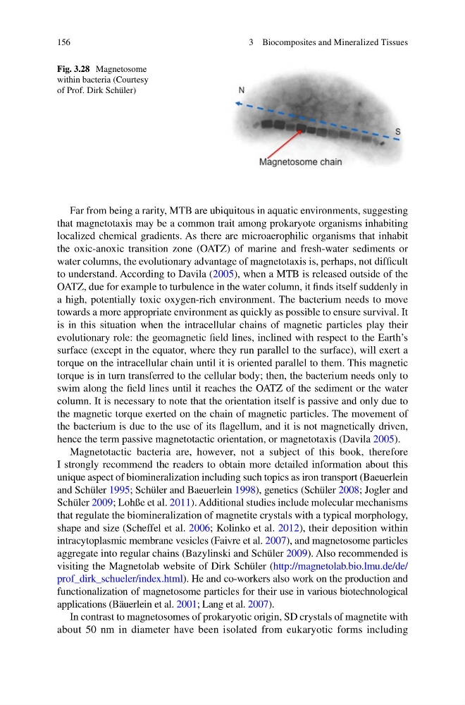
Fig. 3.28 Magnetosome
within bacteria (Courtesy
of Prof. Dirk Schüler)
3 Biocomposites and Mineralized Tissues
Far from being a rarity, MTB are ubiquitous in aquatic environments, suggesting
that magnetotaxis may be a common trait among prokaryote organisms inhabiting
localized chemical gradients. As there are microaerophilic organisms that inhabit
the oxic-anoxic transition zone (OATZ) of marine and fresh-water sediments or
water columns, the evolutionary advantage of magnetotaxis is, perhaps, not diffi cult
to understand. According to Davila ( 2005 ), when a MTB is released outside of the
OATZ, due for example to turbulence in the water column, it fi nds itself suddenly in
a high, potentially toxic oxygen-rich environment. The bacterium needs to move
towards a more appropriate environment as quickly as possible to ensure survival. It
is in this situation when the intracellular chains of magnetic particles play their
evolutionary role: the geomagnetic fi eld lines, inclined with respect to the Earth’s
surface (except in the equator, where they run parallel to the surface), will exert a
torque on the intracellular chain until it is oriented parallel to them. This magnetic
torque is in turn transferred to the cellular body; then, the bacterium needs only to
swim along the fi eld lines until it reaches the OATZ of the sediment or the water
column. It is necessary to note that the orientation itself is passive and only due to
the magnetic torque exerted on the chain of magnetic particles. The movement of
the bacterium is due to the use of its fl agellum, and it is not magnetically driven,
hence the term passive magnetotactic orientation, or magnetotaxis (Davila 2005 ).
Magnetotactic bacteria are, however, not a subject of this book, therefore
I strongly recommend the readers to obtain more detailed information about this
unique aspect of biomineralization including such topics as iron transport (Baeuerlein
and Schüler 1995 ; Schüler and Baeuerlein 1998 ), genetics (Schüler 2008 ; Jogler and
Schüler 2009 ; Lohße et al. 2011 ). Additional studies include molecular mechanisms
that regulate the biomineralization of magnetite crystals with a typical morphology,
shape and size (Scheffel et al. 2006 ; Kolinko et al. 2012 ), their deposition within
intracytoplasmic membrane vesicles (Faivre et al. 2007 ), and magnetosome particles
aggregate into regular chains (Bazylinski and Schüler 2009 ). Also recommended is
visiting the Magnetolab website of Dirk Schüler ( http://magnetolab.bio.lmu.de/de/
prof_dirk_schueler/index.html ). He and co-workers also work on the production and
functionalization of magnetosome particles for their use in various biotechnological
applications (Bäuerlein et al. 2001 ; Lang et al. 2007 ).
In contrast to magnetosomes of prokaryotic origin, SD crystals of magnetite with
about 50 nm in diameter have been isolated from eukaryotic forms including
Fig. 3.28 Magnetosome
within bacteria (Courtesy
of Prof. Dirk Schüler)
3 Biocomposites and Mineralized Tissues