

246
M.L. Cancela et al.
Extracellular proteases, in particular serine proteases, play a major role in
Perkinsus pathogenicity and virulence (La Peyre et al. 1996, Faisal et al. 1999,
Tall et al. 1999). The presence of protease inhibitors in bivalve plasma have been
proposed as a possible mechanism of defense against Perkinsus (Xue et al. 2006).
Thus, inhibitory activity of oyster plasma was weaker in C. virginica than in
C. gigas, which is also less susceptible to P. marinus infection (Faisal et al. 1998).
Furthermore, a negative correlation was reported between disease intensity and
protease inhibitory activity (Oliver et al. 2000).
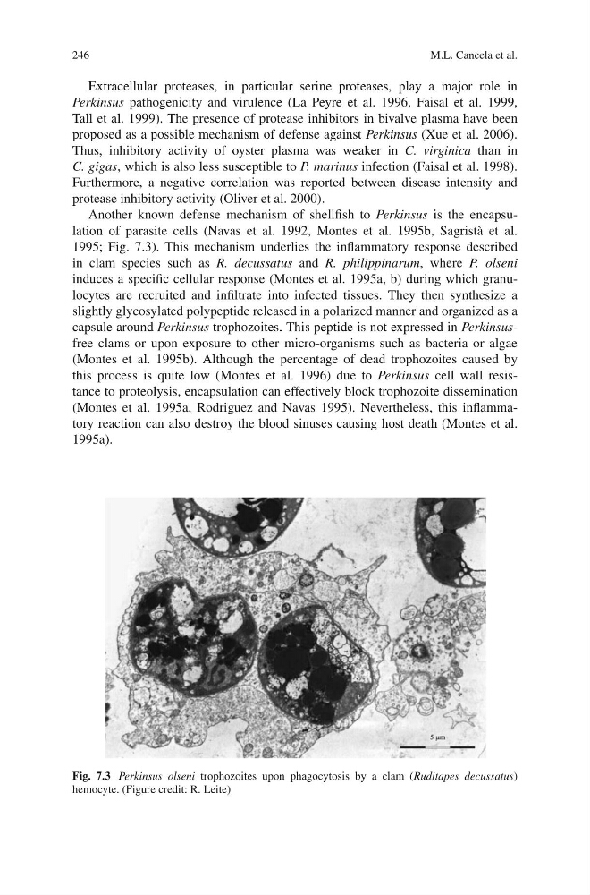
Another known defense mechanism of shellfish to Perkinsus is the encapsulation of parasite cells (Navas et al. 1992, Montes et al. 1995b, Sagristà et al.
1995; Fig. 7.3). This mechanism underlies the inflammatory response described
in clam species such as R. decussatus and R. philippinarum, where P. olseni
induces a specific cellular response (Montes et al. 1995a, b) during which granulocytes are recruited and infiltrate into infected tissues. They then synthesize a
slightly glycosylated polypeptide released in a polarized manner and organized as a
capsule around Perkinsus trophozoites. This peptide is not expressed in Perkinsusfree clams or upon exposure to other micro-organisms such as bacteria or algae
(Montes et al. 1995b). Although the percentage of dead trophozoites caused by
this process is quite low (Montes et al. 1996) due to Perkinsus cell wall resistance to proteolysis, encapsulation can effectively block trophozoite dissemination
(Montes et al. 1995a, Rodriguez and Navas 1995). Nevertheless, this inflammatory reaction can also destroy the blood sinuses causing host death (Montes et al.
1995a).
Fig. 7.3 Perkinsus olseni trophozoites upon phagocytosis by a clam (Ruditapes decussatus)
hemocyte. (Figure credit: R. Leite)
M.L. Cancela et al.
Extracellular proteases, in particular serine proteases, play a major role in
Perkinsus pathogenicity and virulence (La Peyre et al. 1996, Faisal et al. 1999,
Tall et al. 1999). The presence of protease inhibitors in bivalve plasma have been
proposed as a possible mechanism of defense against Perkinsus (Xue et al. 2006).
Thus, inhibitory activity of oyster plasma was weaker in C. virginica than in
C. gigas, which is also less susceptible to P. marinus infection (Faisal et al. 1998).
Furthermore, a negative correlation was reported between disease intensity and
protease inhibitory activity (Oliver et al. 2000).
Another known defense mechanism of shellfish to Perkinsus is the encapsulation of parasite cells (Navas et al. 1992, Montes et al. 1995b, Sagristà et al.
1995; Fig. 7.3). This mechanism underlies the inflammatory response described
in clam species such as R. decussatus and R. philippinarum, where P. olseni
induces a specific cellular response (Montes et al. 1995a, b) during which granulocytes are recruited and infiltrate into infected tissues. They then synthesize a
slightly glycosylated polypeptide released in a polarized manner and organized as a
capsule around Perkinsus trophozoites. This peptide is not expressed in Perkinsusfree clams or upon exposure to other micro-organisms such as bacteria or algae
(Montes et al. 1995b). Although the percentage of dead trophozoites caused by
this process is quite low (Montes et al. 1996) due to Perkinsus cell wall resistance to proteolysis, encapsulation can effectively block trophozoite dissemination
(Montes et al. 1995a, Rodriguez and Navas 1995). Nevertheless, this inflammatory reaction can also destroy the blood sinuses causing host death (Montes et al.
1995a).
Fig. 7.3 Perkinsus olseni trophozoites upon phagocytosis by a clam (Ruditapes decussatus)
hemocyte. (Figure credit: R. Leite)