

ferritins. The turnover rate of ferritin minerals in vivo is not known but studies with
natural maxi-ferritin minerals suggest it could be significant (Castruita et al. 2007).
Dissolution of ferritin minerals has a fast, pH-dependent initial phase and a
slower, pH-independent phase in ~100–200 irons per cage are removed. These iron
atoms may be anchored in the protein and represent iron still in transit through the
proteins (Turano et al. 2010). The phosphate content of ferritin minerals also
influences dissolution of mineral dissolution rates. Rates in the early phase of
mineralization decreases when phosphate is high, whereas the later, slow phase is
independent of phosphate (Richards et al. 1996). Such observations support the
notion that phosphate replaces coordinated water and thereby blocks hydrolysis and
formation ferric oxo bridges, which would have a larger effect on mineral nuclei
than on bulk mineral.
What keeps ferritin minerals dissolving at a rapid rate? The cytoplasm of most
cells is thought to be “reducing,” i.e., full of reductants. Increased rates of iron
mineral dissolution occurred when ferritin threefold pores were unfolded, as
observed in a crystal structure of a ferritin folded normally (Takagi et al. 1998).
Thus, in normal ferritin cages, the folded pores keep reductant away from the ferric
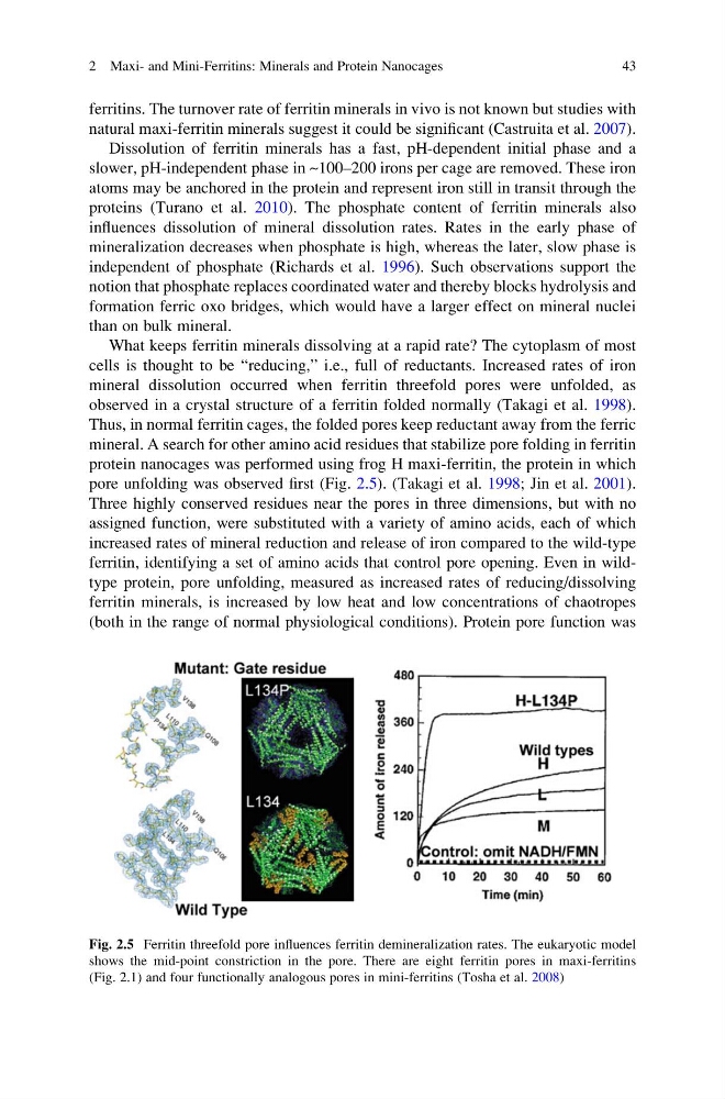
mineral. A search for other amino acid residues that stabilize pore folding in ferritin
protein nanocages was performed using frog H maxi-ferritin, the protein in which
pore unfolding was observed first (Fig. 2.5). (Takagi et al. 1998; Jin et al. 2001).
Three highly conserved residues near the pores in three dimensions, but with no
assigned function, were substituted with a variety of amino acids, each of which
increased rates of mineral reduction and release of iron compared to the wild-type
ferritin, identifying a set of amino acids that control pore opening. Even in wildtype protein, pore unfolding, measured as increased rates of reducing/dissolving
ferritin minerals, is increased by low heat and low concentrations of chaotropes
(both in the range of normal physiological conditions). Protein pore function was
Fig. 2.5 Ferritin threefold pore influences ferritin demineralization rates. The eukaryotic model
shows the mid-point constriction in the pore. There are eight ferritin pores in maxi-ferritins
(Fig. 2.1) and four functionally analogous pores in mini-ferritins (Tosha et al. 2008)
2 Maxi- and Mini-Ferritins: Minerals and Protein Nanocages
43
natural maxi-ferritin minerals suggest it could be significant (Castruita et al. 2007).
Dissolution of ferritin minerals has a fast, pH-dependent initial phase and a
slower, pH-independent phase in ~100–200 irons per cage are removed. These iron
atoms may be anchored in the protein and represent iron still in transit through the
proteins (Turano et al. 2010). The phosphate content of ferritin minerals also
influences dissolution of mineral dissolution rates. Rates in the early phase of
mineralization decreases when phosphate is high, whereas the later, slow phase is
independent of phosphate (Richards et al. 1996). Such observations support the
notion that phosphate replaces coordinated water and thereby blocks hydrolysis and
formation ferric oxo bridges, which would have a larger effect on mineral nuclei
than on bulk mineral.
What keeps ferritin minerals dissolving at a rapid rate? The cytoplasm of most
cells is thought to be “reducing,” i.e., full of reductants. Increased rates of iron
mineral dissolution occurred when ferritin threefold pores were unfolded, as
observed in a crystal structure of a ferritin folded normally (Takagi et al. 1998).
Thus, in normal ferritin cages, the folded pores keep reductant away from the ferric
mineral. A search for other amino acid residues that stabilize pore folding in ferritin
protein nanocages was performed using frog H maxi-ferritin, the protein in which
pore unfolding was observed first (Fig. 2.5). (Takagi et al. 1998; Jin et al. 2001).
Three highly conserved residues near the pores in three dimensions, but with no
assigned function, were substituted with a variety of amino acids, each of which
increased rates of mineral reduction and release of iron compared to the wild-type
ferritin, identifying a set of amino acids that control pore opening. Even in wildtype protein, pore unfolding, measured as increased rates of reducing/dissolving
ferritin minerals, is increased by low heat and low concentrations of chaotropes
(both in the range of normal physiological conditions). Protein pore function was
Fig. 2.5 Ferritin threefold pore influences ferritin demineralization rates. The eukaryotic model
shows the mid-point constriction in the pore. There are eight ferritin pores in maxi-ferritins
(Fig. 2.1) and four functionally analogous pores in mini-ferritins (Tosha et al. 2008)
2 Maxi- and Mini-Ferritins: Minerals and Protein Nanocages
43