

nucleation and mineralization) properties of the minerals, and protein control of
mineral dissolution and release of Fe(II). Pores in ferritin protein cages control iron
entry for mineralization and iron exit after mineral dissolution. The relationship
between phosphate or the presence of catalytically inactive subunits (animal
L subunits) and ferritin iron mineral disorder is developed based on new information about contributions of ferritin protein cage structure to nucleation in protein
cage subunit channels that exit close enough to those of other subunits and exiting
mineral nuclei to facilitate bulk mineral formation. How and where protons move in
and out of the protein during mineral synthesis and dissolution, how ferritin cage
assembly with 12 or 24 subunits is encoded in the widely divergent ferritin amino
acid sequences, and what is the role of the protein in synthesis of the bulk mineral
are all described as problems requiring new approaches in future investigations of
ferritin biominerals.
2.1 Introduction
Ferritins are protein cages that synthesize ferric oxides (hydrated minerals), which
are located in a central cavity of the protein, 5–8 nm in diameter (e.g., [Lewin et al.
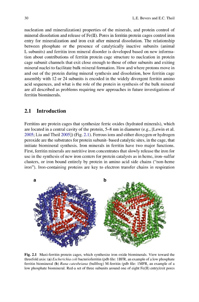
2005; Liu and Theil 2005]) (Fig. 2.1). Ferrous ions and either dioxygen or hydrogen
peroxide are the substrates for protein subunit- based catalytic sites, in the cage, that
initiate biomineral synthesis. Iron minerals in ferritin have two major functions.
First, ferritin minerals are nutritive iron concentrates that slowly release the iron for
use in the synthesis of new iron centers for protein catalysts as in heme, iron–sulfur
clusters, or iron bound entirely by protein in amino acid side chains (“non-heme
iron”). Iron-containing proteins are key to electron transfer chains in respiration
Fig. 2.1 Maxi-ferritin protein cages, which synthesize iron oxide biominerals. View toward the
threefold axis: (a) Escherichia coli bacterioferritin (pdb file: 1BFR, an example of a low phosphate
ferritin biomineral (b) Rana catesbeiana (bullfrog) M-ferritin (pdb file: 1MFR, an example of a
low phosphate biomineral. Red-a set of three subunits around one of eight Fe(II) entry/exit pores
30
L.E. Bevers and E.C. Theil
mineral dissolution and release of Fe(II). Pores in ferritin protein cages control iron
entry for mineralization and iron exit after mineral dissolution. The relationship
between phosphate or the presence of catalytically inactive subunits (animal
L subunits) and ferritin iron mineral disorder is developed based on new information about contributions of ferritin protein cage structure to nucleation in protein
cage subunit channels that exit close enough to those of other subunits and exiting
mineral nuclei to facilitate bulk mineral formation. How and where protons move in
and out of the protein during mineral synthesis and dissolution, how ferritin cage
assembly with 12 or 24 subunits is encoded in the widely divergent ferritin amino
acid sequences, and what is the role of the protein in synthesis of the bulk mineral
are all described as problems requiring new approaches in future investigations of
ferritin biominerals.
2.1 Introduction
Ferritins are protein cages that synthesize ferric oxides (hydrated minerals), which
are located in a central cavity of the protein, 5–8 nm in diameter (e.g., [Lewin et al.
2005; Liu and Theil 2005]) (Fig. 2.1). Ferrous ions and either dioxygen or hydrogen
peroxide are the substrates for protein subunit- based catalytic sites, in the cage, that
initiate biomineral synthesis. Iron minerals in ferritin have two major functions.
First, ferritin minerals are nutritive iron concentrates that slowly release the iron for
use in the synthesis of new iron centers for protein catalysts as in heme, iron–sulfur
clusters, or iron bound entirely by protein in amino acid side chains (“non-heme
iron”). Iron-containing proteins are key to electron transfer chains in respiration
Fig. 2.1 Maxi-ferritin protein cages, which synthesize iron oxide biominerals. View toward the
threefold axis: (a) Escherichia coli bacterioferritin (pdb file: 1BFR, an example of a low phosphate
ferritin biomineral (b) Rana catesbeiana (bullfrog) M-ferritin (pdb file: 1MFR, an example of a
low phosphate biomineral. Red-a set of three subunits around one of eight Fe(II) entry/exit pores
30
L.E. Bevers and E.C. Theil