

and may require external interactions to stabilize its secondary structure (Evans
2008). One of us (P. Narayanappa) works on obtaining the full sequence of
calprismin to confirm this prediction. Other ongoing characterizations include the
Acetic acid-insoluble matrix:
0.5 to 0.6%
Acetic acid-soluble :
0.02 to 0.05%
NACREOUS ARAGONITIC LAYER
a
PROTEIN NAME
Mucoperlin
Apparent MW (kDa)
67
Solubility
Soluble
Theoretical pI
4.9
PTMs
Glycosylations (O-type)
Dominant AA
S, P
Ca-binding
Weak affinity
Other properties
May polymerize (?)
a) Short N-terminus: 11 aa
b) 13 tandem repeats of 31 aa, rich
Primary structure
in S and P: 403 aa
c) C-terminus: acidic & hydrophobic,
3 C in the last 41 aa
Protein family
Mucin-type
Localization
Intertabular
Specific of the nacreous layer
b
c
d
e
f
g
h
i
1
2
3
4
5
6
7
50 m
8 m
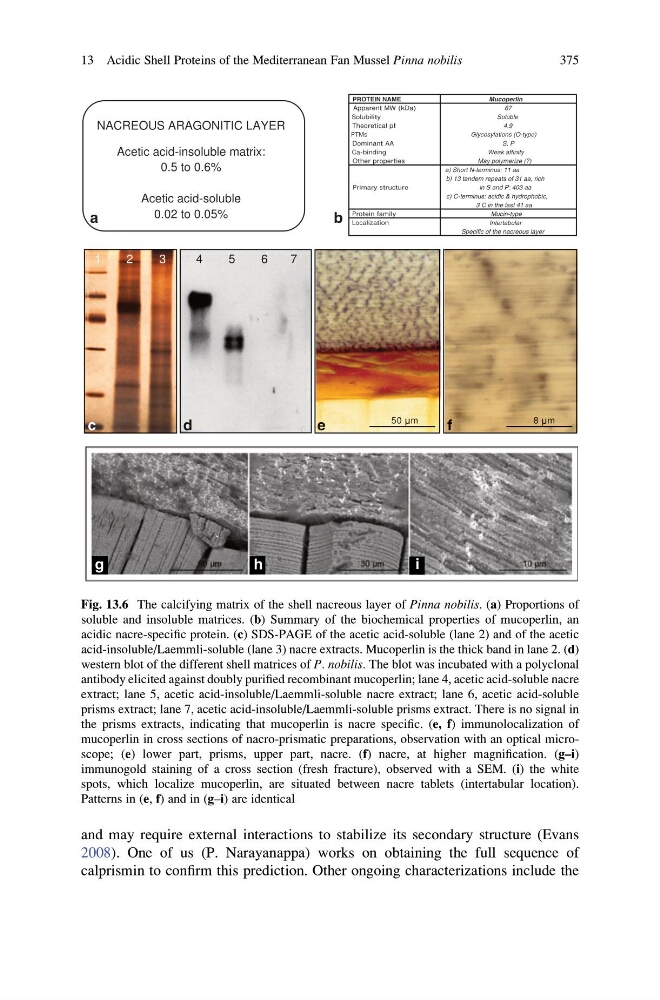
Fig. 13.6 The calcifying matrix of the shell nacreous layer of Pinna nobilis. (a) Proportions of
soluble and insoluble matrices. (b) Summary of the biochemical properties of mucoperlin, an
acidic nacre-specific protein. (c) SDS-PAGE of the acetic acid-soluble (lane 2) and of the acetic
acid-insoluble/Laemmli-soluble (lane 3) nacre extracts. Mucoperlin is the thick band in lane 2. (d)
western blot of the different shell matrices of P. nobilis. The blot was incubated with a polyclonal
antibody elicited against doubly purified recombinant mucoperlin; lane 4, acetic acid-soluble nacre
extract; lane 5, acetic acid-insoluble/Laemmli-soluble nacre extract; lane 6, acetic acid-soluble
prisms extract; lane 7, acetic acid-insoluble/Laemmli-soluble prisms extract. There is no signal in
the prisms extracts, indicating that mucoperlin is nacre specific. (e, f) immunolocalization of
mucoperlin in cross sections of nacro-prismatic preparations, observation with an optical microscope; (e) lower part, prisms, upper part, nacre. (f) nacre, at higher magnification. (g–i)
immunogold staining of a cross section (fresh fracture), observed with a SEM. (i) the white
spots, which localize mucoperlin, are situated between nacre tablets (intertabular location).
Patterns in (e, f) and in (g–i) are identical
13 Acidic Shell Proteins of the Mediterranean Fan Mussel Pinna nobilis
375
2008). One of us (P. Narayanappa) works on obtaining the full sequence of
calprismin to confirm this prediction. Other ongoing characterizations include the
Acetic acid-insoluble matrix:
0.5 to 0.6%
Acetic acid-soluble :
0.02 to 0.05%
NACREOUS ARAGONITIC LAYER
a
PROTEIN NAME
Mucoperlin
Apparent MW (kDa)
67
Solubility
Soluble
Theoretical pI
4.9
PTMs
Glycosylations (O-type)
Dominant AA
S, P
Ca-binding
Weak affinity
Other properties
May polymerize (?)
a) Short N-terminus: 11 aa
b) 13 tandem repeats of 31 aa, rich
Primary structure
in S and P: 403 aa
c) C-terminus: acidic & hydrophobic,
3 C in the last 41 aa
Protein family
Mucin-type
Localization
Intertabular
Specific of the nacreous layer
b
c
d
e
f
g
h
i
1
2
3
4
5
6
7
50 m
8 m
Fig. 13.6 The calcifying matrix of the shell nacreous layer of Pinna nobilis. (a) Proportions of
soluble and insoluble matrices. (b) Summary of the biochemical properties of mucoperlin, an
acidic nacre-specific protein. (c) SDS-PAGE of the acetic acid-soluble (lane 2) and of the acetic
acid-insoluble/Laemmli-soluble (lane 3) nacre extracts. Mucoperlin is the thick band in lane 2. (d)
western blot of the different shell matrices of P. nobilis. The blot was incubated with a polyclonal
antibody elicited against doubly purified recombinant mucoperlin; lane 4, acetic acid-soluble nacre
extract; lane 5, acetic acid-insoluble/Laemmli-soluble nacre extract; lane 6, acetic acid-soluble
prisms extract; lane 7, acetic acid-insoluble/Laemmli-soluble prisms extract. There is no signal in
the prisms extracts, indicating that mucoperlin is nacre specific. (e, f) immunolocalization of
mucoperlin in cross sections of nacro-prismatic preparations, observation with an optical microscope; (e) lower part, prisms, upper part, nacre. (f) nacre, at higher magnification. (g–i)
immunogold staining of a cross section (fresh fracture), observed with a SEM. (i) the white
spots, which localize mucoperlin, are situated between nacre tablets (intertabular location).
Patterns in (e, f) and in (g–i) are identical
13 Acidic Shell Proteins of the Mediterranean Fan Mussel Pinna nobilis
375