

is reduced to 10–20 mm. This intermediate brownish layer is essentially organic and
insoluble. Although of homogeneous aspect once sealed by the subjacent nacreous
layer, it has been shown that this layer results from the coalescence of polygonal
organic elements during its synthesis (Cuif et al. 1985). By many aspects (insolubility, color), the intermediate layer exhibits similarities with the periostracal layer,
and may correspond to chitino-proteinaceous complexes resulting from a quinonetanning process. This layer seals the prismatic layer and represents the template for
60 m
2 0 m
a
c
d
100 m
100 m
10 m
200 m
b
e
f
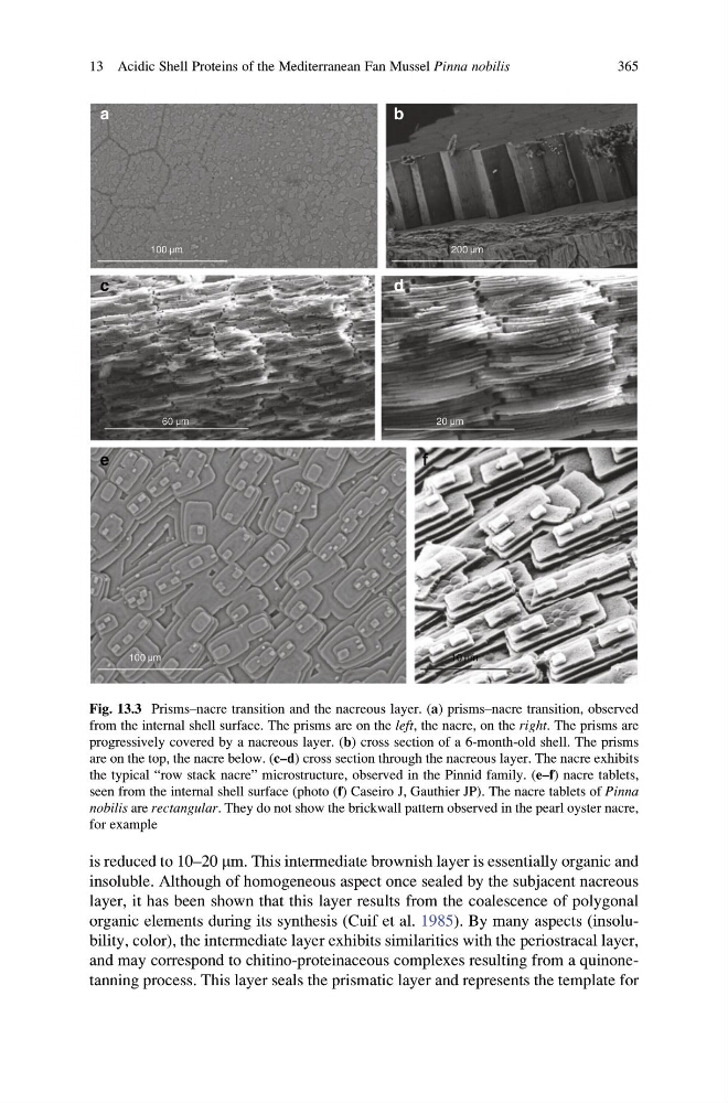
Fig. 13.3 Prisms–nacre transition and the nacreous layer. (a) prisms–nacre transition, observed
from the internal shell surface. The prisms are on the left, the nacre, on the right. The prisms are
progressively covered by a nacreous layer. (b) cross section of a 6-month-old shell. The prisms
are on the top, the nacre below. (c–d) cross section through the nacreous layer. The nacre exhibits
the typical “row stack nacre” microstructure, observed in the Pinnid family. (e–f) nacre tablets,
seen from the internal shell surface (photo (f) Caseiro J, Gauthier JP). The nacre tablets of Pinna
nobilis are rectangular. They do not show the brickwall pattern observed in the pearl oyster nacre,
for example
13 Acidic Shell Proteins of the Mediterranean Fan Mussel Pinna nobilis
365
insoluble. Although of homogeneous aspect once sealed by the subjacent nacreous
layer, it has been shown that this layer results from the coalescence of polygonal
organic elements during its synthesis (Cuif et al. 1985). By many aspects (insolubility, color), the intermediate layer exhibits similarities with the periostracal layer,
and may correspond to chitino-proteinaceous complexes resulting from a quinonetanning process. This layer seals the prismatic layer and represents the template for
60 m
2 0 m
a
c
d
100 m
100 m
10 m
200 m
b
e
f
Fig. 13.3 Prisms–nacre transition and the nacreous layer. (a) prisms–nacre transition, observed
from the internal shell surface. The prisms are on the left, the nacre, on the right. The prisms are
progressively covered by a nacreous layer. (b) cross section of a 6-month-old shell. The prisms
are on the top, the nacre below. (c–d) cross section through the nacreous layer. The nacre exhibits
the typical “row stack nacre” microstructure, observed in the Pinnid family. (e–f) nacre tablets,
seen from the internal shell surface (photo (f) Caseiro J, Gauthier JP). The nacre tablets of Pinna
nobilis are rectangular. They do not show the brickwall pattern observed in the pearl oyster nacre,
for example
13 Acidic Shell Proteins of the Mediterranean Fan Mussel Pinna nobilis
365