

diameters generally increase from the outer to the inner parts of the shell through
geometric selection.” The calcitic prisms of P. nobilis belong to this subcategory.
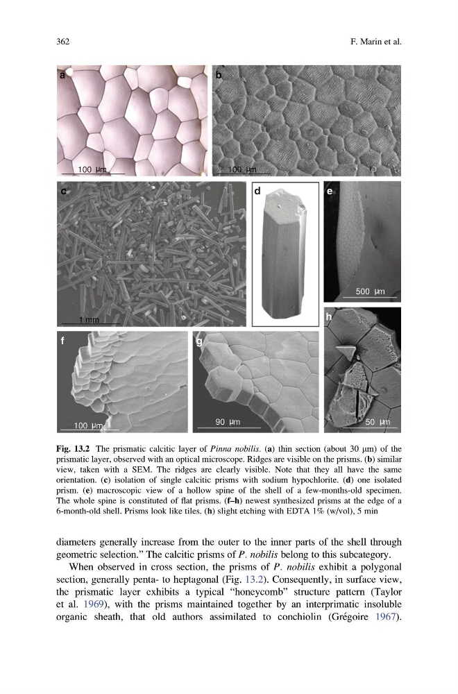
When observed in cross section, the prisms of P. nobilis exhibit a polygonal
section, generally penta- to heptagonal (Fig. 13.2). Consequently, in surface view,
the prismatic layer exhibits a typical “honeycomb” structure pattern (Taylor
et al. 1969), with the prisms maintained together by an interprimatic insoluble
organic sheath, that old authors assimilated to conchiolin (Gre ´goire 1967).
100 m
100 m
1 mm
100 m
90 m
500 m
50 m
a
b
c
d
e
f
g
h
Fig. 13.2 The prismatic calcitic layer of Pinna nobilis. (a) thin section (about 30 mm) of the
prismatic layer, observed with an optical microscope. Ridges are visible on the prisms. (b) similar
view, taken with a SEM. The ridges are clearly visible. Note that they all have the same
orientation. (c) isolation of single calcitic prisms with sodium hypochlorite. (d) one isolated
prism. (e) macroscopic view of a hollow spine of the shell of a few-months-old specimen.
The whole spine is constituted of flat prisms. (f–h) newest synthesized prisms at the edge of a
6-month-old shell. Prisms look like tiles. (h) slight etching with EDTA 1% (w/vol), 5 min
362
F. Marin et al.
geometric selection.” The calcitic prisms of P. nobilis belong to this subcategory.
When observed in cross section, the prisms of P. nobilis exhibit a polygonal
section, generally penta- to heptagonal (Fig. 13.2). Consequently, in surface view,
the prismatic layer exhibits a typical “honeycomb” structure pattern (Taylor
et al. 1969), with the prisms maintained together by an interprimatic insoluble
organic sheath, that old authors assimilated to conchiolin (Gre ´goire 1967).
100 m
100 m
1 mm
100 m
90 m
500 m
50 m
a
b
c
d
e
f
g
h
Fig. 13.2 The prismatic calcitic layer of Pinna nobilis. (a) thin section (about 30 mm) of the
prismatic layer, observed with an optical microscope. Ridges are visible on the prisms. (b) similar
view, taken with a SEM. The ridges are clearly visible. Note that they all have the same
orientation. (c) isolation of single calcitic prisms with sodium hypochlorite. (d) one isolated
prism. (e) macroscopic view of a hollow spine of the shell of a few-months-old specimen.
The whole spine is constituted of flat prisms. (f–h) newest synthesized prisms at the edge of a
6-month-old shell. Prisms look like tiles. (h) slight etching with EDTA 1% (w/vol), 5 min
362
F. Marin et al.