

populations showed that the growth kinetics is submitted to slight variations
(Garcia-March et al. 2002; Rabaoui et al. 2007), according to environmental
parameters, including temperatures, water turbidity, depth, nutrient fluxes.
Because the growth of the shell is rapid during the first years (a daily growth
above 0.3 mm for a 1-year-old specimen), it is likely that juvenile specimens
allocate an important part of their energetic budget to the calcification of their
shell, which, in other words, may mean that other metabolic activities are kept low.
However, to our knowledge, there are no precise data in the literature on this aspect
of the physiology of Pinna nobilis.
The shells of young specimens are characterized by the formation of thin calcitic
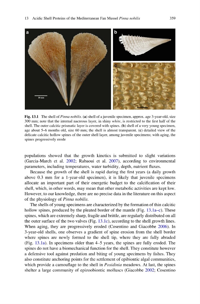
hollow spines, produced by the pleated border of the mantle (Fig. 13.1a–c). These
spines, which are extremely sharp, fragile and brittle, are regularly distributed on all
the outer surface of the two valves (Fig. 13.1c), according to the shell growth lines.
When aging, they are progressively eroded (Cosentino and Giacobbe 2006). In
3-year-old shells, one observes a gradient of spine erosion from the shell border
where spines are newly formed to the shell tip, where they are fully abraded
(Fig. 13.1a). In specimens older than 4–5 years, the spines are fully eroded. The
spines do not have a biomechanical function for the shell. They constitute however
a defensive tool against predation and biting of young specimens by fishes. They
also constitute anchoring points for the settlement of epibiontic algal communities,
which provide a camouflage to the shell in Posidinia meadows. At last, the spines
shelter a large community of epizoobiontic molluscs (Giacobbe 2002; Cosentino
Fig. 13.1 The shell of Pinna nobilis. (a) shell of a juvenile specimen, approx. age 3-year-old, size
300 mm; note that the internal nacreous layer, in shiny white, is restricted to the first half of the
shell. The outer calcitic prismatic layer is covered with spines. (b) shell of a very young specimen,
age about 5–6 months old, size 60 mm; the shell is almost transparent. (c) detailed view of the
delicate calcitic hollow spines of the outer shell layer, among juvenile specimens; with aging, the
spines progressively erode
13 Acidic Shell Proteins of the Mediterranean Fan Mussel Pinna nobilis
359
(Garcia-March et al. 2002; Rabaoui et al. 2007), according to environmental
parameters, including temperatures, water turbidity, depth, nutrient fluxes.
Because the growth of the shell is rapid during the first years (a daily growth
above 0.3 mm for a 1-year-old specimen), it is likely that juvenile specimens
allocate an important part of their energetic budget to the calcification of their
shell, which, in other words, may mean that other metabolic activities are kept low.
However, to our knowledge, there are no precise data in the literature on this aspect
of the physiology of Pinna nobilis.
The shells of young specimens are characterized by the formation of thin calcitic
hollow spines, produced by the pleated border of the mantle (Fig. 13.1a–c). These
spines, which are extremely sharp, fragile and brittle, are regularly distributed on all
the outer surface of the two valves (Fig. 13.1c), according to the shell growth lines.
When aging, they are progressively eroded (Cosentino and Giacobbe 2006). In
3-year-old shells, one observes a gradient of spine erosion from the shell border
where spines are newly formed to the shell tip, where they are fully abraded
(Fig. 13.1a). In specimens older than 4–5 years, the spines are fully eroded. The
spines do not have a biomechanical function for the shell. They constitute however
a defensive tool against predation and biting of young specimens by fishes. They
also constitute anchoring points for the settlement of epibiontic algal communities,
which provide a camouflage to the shell in Posidinia meadows. At last, the spines
shelter a large community of epizoobiontic molluscs (Giacobbe 2002; Cosentino
Fig. 13.1 The shell of Pinna nobilis. (a) shell of a juvenile specimen, approx. age 3-year-old, size
300 mm; note that the internal nacreous layer, in shiny white, is restricted to the first half of the
shell. The outer calcitic prismatic layer is covered with spines. (b) shell of a very young specimen,
age about 5–6 months old, size 60 mm; the shell is almost transparent. (c) detailed view of the
delicate calcitic hollow spines of the outer shell layer, among juvenile specimens; with aging, the
spines progressively erode
13 Acidic Shell Proteins of the Mediterranean Fan Mussel Pinna nobilis
359