

(six silicatein-a isoenzymes; Kaluzhnaya et al. 2005; Wiens et al. 2006), and the
hexactinellid sponges, Crateromorpha meyeri (M€ uller et al. 2008c) and M. chuni
(M€ uller et al. 2009a). The recombinant proteins can be prepared using both
prokaryotic (Escherichia coli) and eukaryotic (Pichia pastori) systems (bioreactor/
laboratory scale). The purified proteins can be applied for the biocatalytic formation
of amorphous silica (biosilica) from monomeric precursors at mild (room temperature, near-neutral pH, aqueous buffer systems) conditions (Schr€ oder et al. 2008).
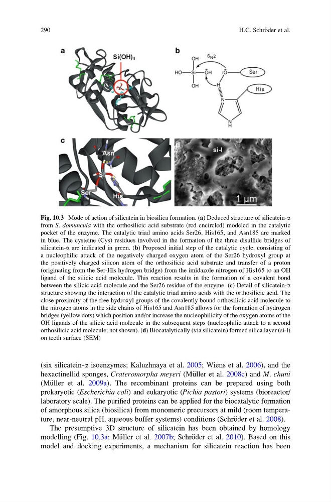
The presumptive 3D structure of silicatein has been obtained by homology
modelling (Fig. 10.3a; M€ uller et al. 2007b; Schr€ oder et al. 2010). Based on this
model and docking experiments, a mechanism for silicatein reaction has been
Fig. 10.3 Mode of action of silicatein in biosilica formation. (a) Deduced structure of silicatein-a
from S. domuncula with the orthosilicic acid substrate (red encircled) modeled in the catalytic
pocket of the enzyme. The catalytic triad amino acids Ser26, His165, and Asn185 are marked
in blue. The cysteine (Cys) residues involved in the formation of the three disulfide bridges of
silicatein-a are indicated in green. (b) Proposed initial step of the catalytic cycle, consisting of
a nucleophilic attack of the negatively charged oxygen atom of the Ser26 hydroxyl group at
the positively charged silicon atom of the orthosilicic acid substrate and transfer of a proton
(originating from the Ser-His hydrogen bridge) from the imidazole nitrogen of His165 to an OH
ligand of the silicic acid molecule. This reaction results in the formation of a covalent bond
between the silicic acid molecule and the Ser26 residue of the enzyme. (c) Detail of silicatein-a
structure showing the interaction of the catalytic triad amino acids with the orthosilicic acid. The
close proximity of the free hydroxyl groups of the covalently bound orthosilicic acid molecule to
the nitrogen atoms in the side chains of His165 and Asn185 allows for the formation of hydrogen
bridges (yellow dots) which position and/or increase the nucleophilicity of the oxygen atoms of the
OH ligands of the silicic acid molecule in the subsequent steps (nucleophilic attack to a second
orthosilicic acid molecule; not shown). (d) Biocatalytically (via silicatein) formed silica layer (si-l)
on teeth surface (SEM)
290
H.C. Schr€ oder et al.
hexactinellid sponges, Crateromorpha meyeri (M€ uller et al. 2008c) and M. chuni
(M€ uller et al. 2009a). The recombinant proteins can be prepared using both
prokaryotic (Escherichia coli) and eukaryotic (Pichia pastori) systems (bioreactor/
laboratory scale). The purified proteins can be applied for the biocatalytic formation
of amorphous silica (biosilica) from monomeric precursors at mild (room temperature, near-neutral pH, aqueous buffer systems) conditions (Schr€ oder et al. 2008).
The presumptive 3D structure of silicatein has been obtained by homology
modelling (Fig. 10.3a; M€ uller et al. 2007b; Schr€ oder et al. 2010). Based on this
model and docking experiments, a mechanism for silicatein reaction has been
Fig. 10.3 Mode of action of silicatein in biosilica formation. (a) Deduced structure of silicatein-a
from S. domuncula with the orthosilicic acid substrate (red encircled) modeled in the catalytic
pocket of the enzyme. The catalytic triad amino acids Ser26, His165, and Asn185 are marked
in blue. The cysteine (Cys) residues involved in the formation of the three disulfide bridges of
silicatein-a are indicated in green. (b) Proposed initial step of the catalytic cycle, consisting of
a nucleophilic attack of the negatively charged oxygen atom of the Ser26 hydroxyl group at
the positively charged silicon atom of the orthosilicic acid substrate and transfer of a proton
(originating from the Ser-His hydrogen bridge) from the imidazole nitrogen of His165 to an OH
ligand of the silicic acid molecule. This reaction results in the formation of a covalent bond
between the silicic acid molecule and the Ser26 residue of the enzyme. (c) Detail of silicatein-a
structure showing the interaction of the catalytic triad amino acids with the orthosilicic acid. The
close proximity of the free hydroxyl groups of the covalently bound orthosilicic acid molecule to
the nitrogen atoms in the side chains of His165 and Asn185 allows for the formation of hydrogen
bridges (yellow dots) which position and/or increase the nucleophilicity of the oxygen atoms of the
OH ligands of the silicic acid molecule in the subsequent steps (nucleophilic attack to a second
orthosilicic acid molecule; not shown). (d) Biocatalytically (via silicatein) formed silica layer (si-l)
on teeth surface (SEM)
290
H.C. Schr€ oder et al.