

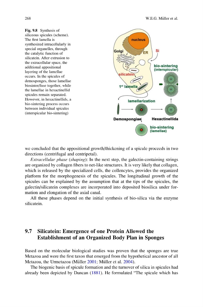
we concluded that the appositional growth/thickening of a spicule proceeds in two
directions (centrifugal and centripetal).
Extracellular phase (shaping): In the next step, the galectin-containing strings
are organized by collagen fibers to net-like structures. It is very likely that collagen,
which is released by the specialized cells, the collencytes, provides the organized
platform for the morphogenesis of the spicules. The longitudinal growth of the
spicules can be explained by the assumption that at the tips of the spicules, the
galectin/silicatein complexes are incorporated into deposited biosilica under formation and elongation of the axial canal.
All these phases depend on the initial synthesis of bio-silica via the enzyme
silicatein.
9.7 Silicatein: Emergence of one Protein Allowed the
Establishment of an Organized Body Plan in Sponges
Based on the molecular biological studies was proven that the sponges are true
Metazoa and were the first taxon that emerged from the hypothetical ancestor of all
Metazoa, the Urmetazoa (M€ uller 2001; M€ uller et al. 2004).
The biogenic basis of spicule formation and the turnover of silica in spicules had
already been depicted by Duncan (1881). He formulated “The spicule which has
Fig. 9.8 Synthesis of
siliceous spicules (scheme).
The first lamella is
synthesized intracellularly in
special organelles, through
the catalytic function of
silicatein. After extrusion to
the extracellular space, the
additional appositional
layering of the lamellae
occurs. In the spicules of
demosponges, those lamellae
biosinter/fuse together, while
the lamellae in hexactinellid
spicules remain separated.
However, in hexactinellids, a
bio-sintering process occurs
between individual spicules
(interspicular bio-sintering)
268
W.E.G. M€ uller et al.
directions (centrifugal and centripetal).
Extracellular phase (shaping): In the next step, the galectin-containing strings
are organized by collagen fibers to net-like structures. It is very likely that collagen,
which is released by the specialized cells, the collencytes, provides the organized
platform for the morphogenesis of the spicules. The longitudinal growth of the
spicules can be explained by the assumption that at the tips of the spicules, the
galectin/silicatein complexes are incorporated into deposited biosilica under formation and elongation of the axial canal.
All these phases depend on the initial synthesis of bio-silica via the enzyme
silicatein.
9.7 Silicatein: Emergence of one Protein Allowed the
Establishment of an Organized Body Plan in Sponges
Based on the molecular biological studies was proven that the sponges are true
Metazoa and were the first taxon that emerged from the hypothetical ancestor of all
Metazoa, the Urmetazoa (M€ uller 2001; M€ uller et al. 2004).
The biogenic basis of spicule formation and the turnover of silica in spicules had
already been depicted by Duncan (1881). He formulated “The spicule which has
Fig. 9.8 Synthesis of
siliceous spicules (scheme).
The first lamella is
synthesized intracellularly in
special organelles, through
the catalytic function of
silicatein. After extrusion to
the extracellular space, the
additional appositional
layering of the lamellae
occurs. In the spicules of
demosponges, those lamellae
biosinter/fuse together, while
the lamellae in hexactinellid
spicules remain separated.
However, in hexactinellids, a
bio-sintering process occurs
between individual spicules
(interspicular bio-sintering)
268
W.E.G. M€ uller et al.