

proteins levels, mis-regulation in gene expression and DNA damage (Batel et al.
1998; Schr€ oder et al. 2005; Bonaventura et al. 2005, 2006; Holzinger and L€ utz
2006; Tedetti and Sempe ´re ´ 2007; Banaszak and Lesser 2009). In the following, we
will briefly review the effects of ionizing radiation on the formation of the skeleton.
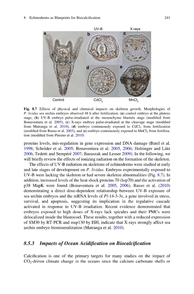
The effects of UV-B radiation on skeletons of echinoderms were studied at early
and late stages of development on P. lividus. Embryos experimentally exposed to
UV-B were lacking the skeleton or had severe skeleton abnormalities (Fig. 8.7). In
addition, increased levels of the heat shock proteins 70 (hsp70) and the activation of
p38 MapK were found (Bonaventura et al. 2005, 2006). Russo et al. (2010)
demonstrating a direct dose-dependent relationship between UV-B exposure of
sea urchin embryos and the mRNA levels of Pl-14-3-3e, a gene involved in stress,
survival, and apoptosis, suggesting its implication in the regulative cascade
activated in response to UV-B irradiation. Recent evidence demonstrated that
embryos exposed to high doses of X-rays lack spicules and their PMCs were
delocalized inside the blastocoel. These results, together with a reduced expression
of SM30 by RT-PCR and msp130 by ISH, indicate that X-rays strongly affect sea
urchin embryo biomineralization (Matranga et al. 2010).
8.5.3 Impacts of Ocean Acidification on Biocalcification
Calcification is one of the primary targets for many studies on the impact of
CO 2 -driven climate change in the oceans since the calcium carbonate shells or
Fig. 8.7 Effects of physical and chemical impacts on skeleton growth. Morphologies of
P. lividus sea urchin embryos observed 48 h after fertilization. (a) control embryo at the pluteus
stage, (b) UV-B embryo pulse-irradiated at the mesenchyme blastula stage (modified from
Bonaventura et al. 2005), (c) X-rays embryo pulse-irradiated at the cleavage stage (modified
from Matranga et al. 2010), (d) embryo continuously exposed to CdCl 2 from fertilization
(modified from Russo et al. 2003), and (e) embryo continuously exposed to MnCl 2 from fertilization (modified from Pinsino et al. 2010)
8 Echinoderms as Blueprints for Biocalcification
241
1998; Schr€ oder et al. 2005; Bonaventura et al. 2005, 2006; Holzinger and L€ utz
2006; Tedetti and Sempe ´re ´ 2007; Banaszak and Lesser 2009). In the following, we
will briefly review the effects of ionizing radiation on the formation of the skeleton.
The effects of UV-B radiation on skeletons of echinoderms were studied at early
and late stages of development on P. lividus. Embryos experimentally exposed to
UV-B were lacking the skeleton or had severe skeleton abnormalities (Fig. 8.7). In
addition, increased levels of the heat shock proteins 70 (hsp70) and the activation of
p38 MapK were found (Bonaventura et al. 2005, 2006). Russo et al. (2010)
demonstrating a direct dose-dependent relationship between UV-B exposure of
sea urchin embryos and the mRNA levels of Pl-14-3-3e, a gene involved in stress,
survival, and apoptosis, suggesting its implication in the regulative cascade
activated in response to UV-B irradiation. Recent evidence demonstrated that
embryos exposed to high doses of X-rays lack spicules and their PMCs were
delocalized inside the blastocoel. These results, together with a reduced expression
of SM30 by RT-PCR and msp130 by ISH, indicate that X-rays strongly affect sea
urchin embryo biomineralization (Matranga et al. 2010).
8.5.3 Impacts of Ocean Acidification on Biocalcification
Calcification is one of the primary targets for many studies on the impact of
CO 2 -driven climate change in the oceans since the calcium carbonate shells or
Fig. 8.7 Effects of physical and chemical impacts on skeleton growth. Morphologies of
P. lividus sea urchin embryos observed 48 h after fertilization. (a) control embryo at the pluteus
stage, (b) UV-B embryo pulse-irradiated at the mesenchyme blastula stage (modified from
Bonaventura et al. 2005), (c) X-rays embryo pulse-irradiated at the cleavage stage (modified
from Matranga et al. 2010), (d) embryo continuously exposed to CdCl 2 from fertilization
(modified from Russo et al. 2003), and (e) embryo continuously exposed to MnCl 2 from fertilization (modified from Pinsino et al. 2010)
8 Echinoderms as Blueprints for Biocalcification
241