

membrane trafficking. After the initial precipitation occurring inside the cells,
crystals are secreted to the extracellular blastocoelic environment, by fusion of
vesicles with the plasma membrane, where they interact with matrix proteins to
acquire the final crystal form (Wilt et al. 2008; Yang et al. 2011). Thus, the mineral
is believed to be enrobed by the syncytial membrane and the cytoplasm.
The larval skeleton displays considerable morphological diversity among sea
urchin species, including variations in the number, structure/shape, and size of rods.
Thus, when describing the single parts of the skeleton, one should refer to the
species taken into consideration. Here, we want to briefly describe for the first
time the development of the skeleton of P. lividus sea urchin embryo, identifying
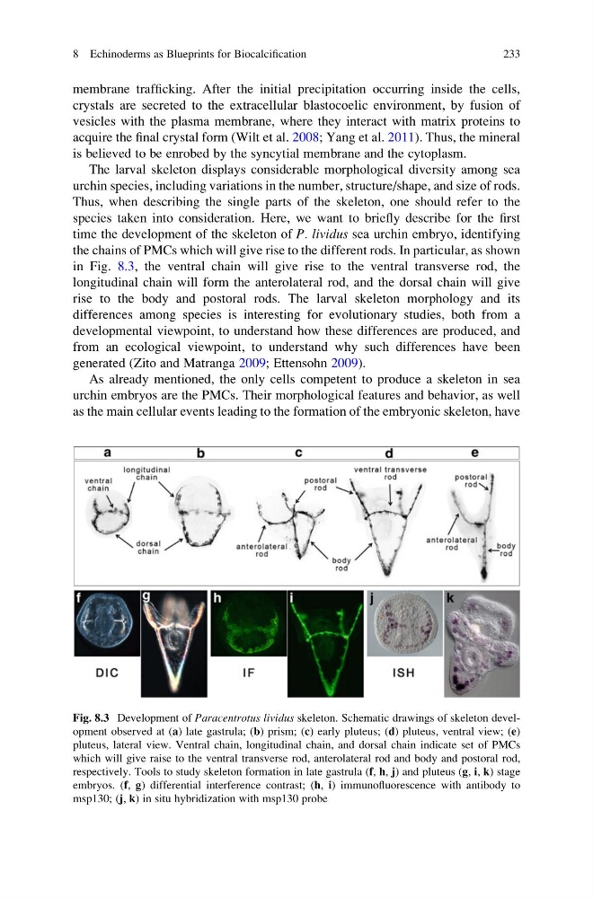
the chains of PMCs which will give rise to the different rods. In particular, as shown
in Fig. 8.3, the ventral chain will give rise to the ventral transverse rod, the
longitudinal chain will form the anterolateral rod, and the dorsal chain will give
rise to the body and postoral rods. The larval skeleton morphology and its
differences among species is interesting for evolutionary studies, both from a
developmental viewpoint, to understand how these differences are produced, and
from an ecological viewpoint, to understand why such differences have been
generated (Zito and Matranga 2009; Ettensohn 2009).
As already mentioned, the only cells competent to produce a skeleton in sea
urchin embryos are the PMCs. Their morphological features and behavior, as well
as the main cellular events leading to the formation of the embryonic skeleton, have
Fig. 8.3 Development of Paracentrotus lividus skeleton. Schematic drawings of skeleton development observed at (a) late gastrula; (b) prism; (c) early pluteus; (d) pluteus, ventral view; (e)
pluteus, lateral view. Ventral chain, longitudinal chain, and dorsal chain indicate set of PMCs
which will give raise to the ventral transverse rod, anterolateral rod and body and postoral rod,
respectively. Tools to study skeleton formation in late gastrula (f, h, j) and pluteus (g, i, k) stage
embryos. (f, g) differential interference contrast; (h, i) immunofluorescence with antibody to
msp130; (j, k) in situ hybridization with msp130 probe
8 Echinoderms as Blueprints for Biocalcification
233
crystals are secreted to the extracellular blastocoelic environment, by fusion of
vesicles with the plasma membrane, where they interact with matrix proteins to
acquire the final crystal form (Wilt et al. 2008; Yang et al. 2011). Thus, the mineral
is believed to be enrobed by the syncytial membrane and the cytoplasm.
The larval skeleton displays considerable morphological diversity among sea
urchin species, including variations in the number, structure/shape, and size of rods.
Thus, when describing the single parts of the skeleton, one should refer to the
species taken into consideration. Here, we want to briefly describe for the first
time the development of the skeleton of P. lividus sea urchin embryo, identifying
the chains of PMCs which will give rise to the different rods. In particular, as shown
in Fig. 8.3, the ventral chain will give rise to the ventral transverse rod, the
longitudinal chain will form the anterolateral rod, and the dorsal chain will give
rise to the body and postoral rods. The larval skeleton morphology and its
differences among species is interesting for evolutionary studies, both from a
developmental viewpoint, to understand how these differences are produced, and
from an ecological viewpoint, to understand why such differences have been
generated (Zito and Matranga 2009; Ettensohn 2009).
As already mentioned, the only cells competent to produce a skeleton in sea
urchin embryos are the PMCs. Their morphological features and behavior, as well
as the main cellular events leading to the formation of the embryonic skeleton, have
Fig. 8.3 Development of Paracentrotus lividus skeleton. Schematic drawings of skeleton development observed at (a) late gastrula; (b) prism; (c) early pluteus; (d) pluteus, ventral view; (e)
pluteus, lateral view. Ventral chain, longitudinal chain, and dorsal chain indicate set of PMCs
which will give raise to the ventral transverse rod, anterolateral rod and body and postoral rod,
respectively. Tools to study skeleton formation in late gastrula (f, h, j) and pluteus (g, i, k) stage
embryos. (f, g) differential interference contrast; (h, i) immunofluorescence with antibody to
msp130; (j, k) in situ hybridization with msp130 probe
8 Echinoderms as Blueprints for Biocalcification
233