

ferrous iron and thus a change in the complex-binding constant or decomposition
of the siderophore. M. magnetotacticum (Paoletti and Blakemore 1986), MV-1
(Dubbels et al. 2004) and M. magneticum have been reported to secrete
siderophores in amounts dependent on the medium iron concentrations and cellular
demand. The latter species was shown to secrete hydroxamates and the catechol
siderophore 3,4-dihydroxybenzoic acid for Fe
III complexation (Calugay et al. 2003;
Calugay et al. 2006). M. magnetotacticum has been reported to show unusual
siderophore production behavior, as production increased with higher ferric iron
concentrations (Paoletti and Blakemore 1986) instead of downregulation when
supply is high. It has been speculated, however, that very rapid assimilation of
dissolved iron and the subsequent low concentration levels triggering siderophore
production might account for this effect (Calugay et al. 2003; Calugay et al. 2004).
Surprisingly, no siderophore production has been detected in cultures of
M. gryphiswaldense although spent media fluid increased ferric iron uptake
which was also shown to be energy coupled in this organism and might indicate
the existence of other potential chelators involved in ferric iron uptake. Ferrous iron
acquisition seemed to be diffusion based and not coupled to energetically driven
uptake (Sch€ uler and Baeuerlein 1996; Sch€ uler and Baeuerlein 1998). Thus, a
general role of siderophores related to iron uptake necessary for magnetosome
formation is still debated and might be species dependent.
No mechanism common to all MTB has been revealed yet, but a number of
proteins involved in general iron uptake have been identified. It is not clear whether
these proteins are part of biochemical pathways involved in biomineralization or in
simply providing essential iron for various metabolic processes. In MV-1, a major
OM
F e (i n )
H+(out)
Magnetosome
Filament
CM
Anchor
Fe(in)
Fe(II)/(III)
Fe(in)
Fe(in)
a
b
c
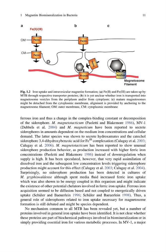
Fig. 1.2 Iron uptake and intravesicular magnetite formation; (a) Fe(II) and Fe(III) are taken up by
MTB through respective transporter proteins; (b) it is yet unclear whether iron is transported into
magnetosome vesicles from the periplasm and/or from cytoplasm; (c) mature magnetosomes
might be detached from the cytoplasmic membrane, alignment is provided by anchoring to the
magnetosome filament; OM: outer membrane, CM: cytoplasmic membrane
1 Magnetite Biomineralization in Bacteria
11
of the siderophore. M. magnetotacticum (Paoletti and Blakemore 1986), MV-1
(Dubbels et al. 2004) and M. magneticum have been reported to secrete
siderophores in amounts dependent on the medium iron concentrations and cellular
demand. The latter species was shown to secrete hydroxamates and the catechol
siderophore 3,4-dihydroxybenzoic acid for Fe
III complexation (Calugay et al. 2003;
Calugay et al. 2006). M. magnetotacticum has been reported to show unusual
siderophore production behavior, as production increased with higher ferric iron
concentrations (Paoletti and Blakemore 1986) instead of downregulation when
supply is high. It has been speculated, however, that very rapid assimilation of
dissolved iron and the subsequent low concentration levels triggering siderophore
production might account for this effect (Calugay et al. 2003; Calugay et al. 2004).
Surprisingly, no siderophore production has been detected in cultures of
M. gryphiswaldense although spent media fluid increased ferric iron uptake
which was also shown to be energy coupled in this organism and might indicate
the existence of other potential chelators involved in ferric iron uptake. Ferrous iron
acquisition seemed to be diffusion based and not coupled to energetically driven
uptake (Sch€ uler and Baeuerlein 1996; Sch€ uler and Baeuerlein 1998). Thus, a
general role of siderophores related to iron uptake necessary for magnetosome
formation is still debated and might be species dependent.
No mechanism common to all MTB has been revealed yet, but a number of
proteins involved in general iron uptake have been identified. It is not clear whether
these proteins are part of biochemical pathways involved in biomineralization or in
simply providing essential iron for various metabolic processes. In MV-1, a major
OM
F e (i n )
H+(out)
Magnetosome
Filament
CM
Anchor
Fe(in)
Fe(II)/(III)
Fe(in)
Fe(in)
a
b
c
Fig. 1.2 Iron uptake and intravesicular magnetite formation; (a) Fe(II) and Fe(III) are taken up by
MTB through respective transporter proteins; (b) it is yet unclear whether iron is transported into
magnetosome vesicles from the periplasm and/or from cytoplasm; (c) mature magnetosomes
might be detached from the cytoplasmic membrane, alignment is provided by anchoring to the
magnetosome filament; OM: outer membrane, CM: cytoplasmic membrane
1 Magnetite Biomineralization in Bacteria
11