

7.2.5 Recent Work on the Structure and Composition
of the Embryonic Spicule
The gross morphology of the endoskeletal spicule in the pluteus stage is a speciesspecific character. The location of branches, small spurs, and fenestrations is
reproducible and specific. Okazaki and Inoue (1976) carried out pioneering work
on the orientation of the crystal axes, and showed that the founding mineral granule
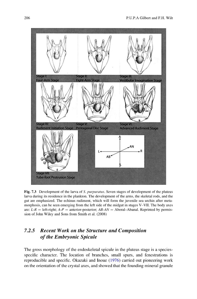
Fig. 7.3 Development of the larva of S. purpuratus. Seven stages of development of the pluteus
larva during its residence in the plankton. The development of the arms, the skeletal rods, and the
gut are emphasized. The echinus rudiment, which will form the juvenile sea urchin after metamorphosis, can be seen emerging from the left side of the midgut in stages V–VII. The body axes
are: L-R ¼ left-right; A-P ¼ anterior-posterior; AB-AN ¼ Aboral–Abanal. Reprinted by permission of John Wiley and Sons from Smith et al. (2008)
206
P.U.P.A Gilbert and F.H. Wilt
of the Embryonic Spicule
The gross morphology of the endoskeletal spicule in the pluteus stage is a speciesspecific character. The location of branches, small spurs, and fenestrations is
reproducible and specific. Okazaki and Inoue (1976) carried out pioneering work
on the orientation of the crystal axes, and showed that the founding mineral granule
Fig. 7.3 Development of the larva of S. purpuratus. Seven stages of development of the pluteus
larva during its residence in the plankton. The development of the arms, the skeletal rods, and the
gut are emphasized. The echinus rudiment, which will form the juvenile sea urchin after metamorphosis, can be seen emerging from the left side of the midgut in stages V–VII. The body axes
are: L-R ¼ left-right; A-P ¼ anterior-posterior; AB-AN ¼ Aboral–Abanal. Reprinted by permission of John Wiley and Sons from Smith et al. (2008)
206
P.U.P.A Gilbert and F.H. Wilt