

the lapillus hierarchical structure. The aragonite fibrils in the sagittae are all
perpendicular to the daily increment layer so that the section of sagitta seems to
be radial. Figure 6.7d shows clearly the asymmetrical location of the nucleus, which
is close to the sulcate side (the left side in Fig. 6.7d). The unusual shape of the
otolith is partly explained by the distribution of the epithelium cells, and particularly the sensory epithelium.
The discovery of the hierarchical structure of the carp otolith can help us to
investigate the biologic self-assembling mechanism and the relationship between
the microstructure and the physiological function of the otolith. The hierarchical
structure of the otoliths can give us more information for investigating the fish
ecology and the hydrochemistry. The particular crystal array of otoliths can also
provide the inspiration on biomimetic and synthetic materials.
6.2.2 Hierarchical Structure of Nacreous Layer in Mollusc Shells
Oaki and Imai (2005) investigated the nacreous layer of Japanese pearl oyster:
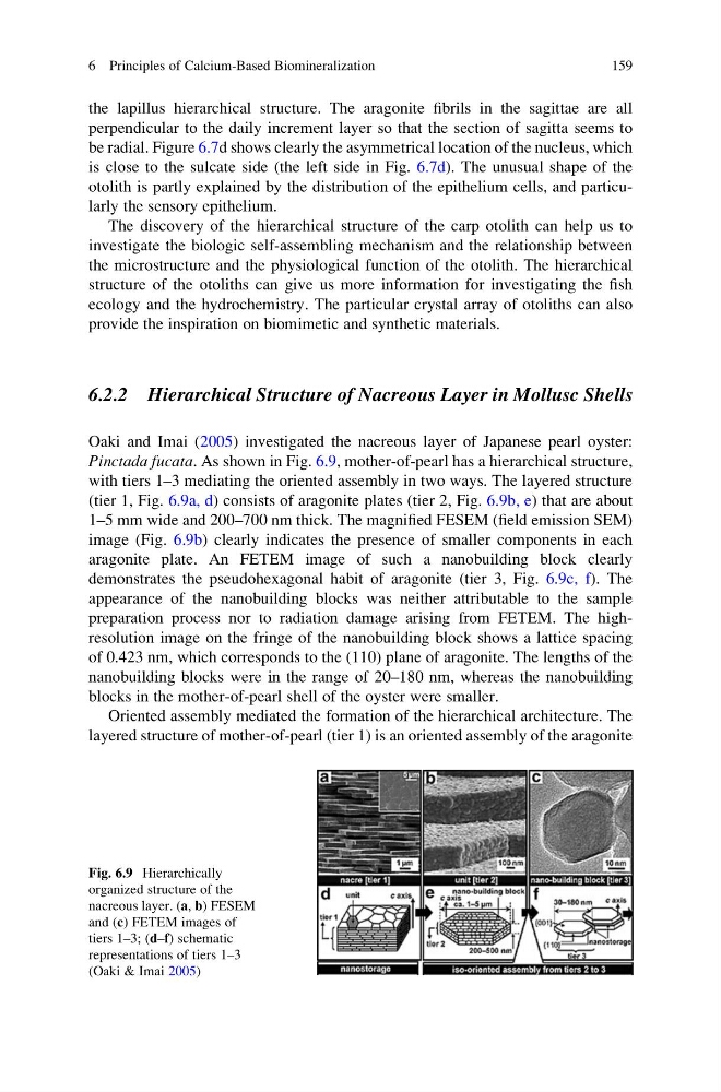
Pinctada fucata. As shown in Fig. 6.9, mother-of-pearl has a hierarchical structure,
with tiers 1–3 mediating the oriented assembly in two ways. The layered structure
(tier 1, Fig. 6.9a, d) consists of aragonite plates (tier 2, Fig. 6.9b, e) that are about
1–5 mm wide and 200–700 nm thick. The magnified FESEM (field emission SEM)
image (Fig. 6.9b) clearly indicates the presence of smaller components in each
aragonite plate. An FETEM image of such a nanobuilding block clearly
demonstrates the pseudohexagonal habit of aragonite (tier 3, Fig. 6.9c, f). The
appearance of the nanobuilding blocks was neither attributable to the sample
preparation process nor to radiation damage arising from FETEM. The highresolution image on the fringe of the nanobuilding block shows a lattice spacing
of 0.423 nm, which corresponds to the (110) plane of aragonite. The lengths of the
nanobuilding blocks were in the range of 20–180 nm, whereas the nanobuilding
blocks in the mother-of-pearl shell of the oyster were smaller.
Oriented assembly mediated the formation of the hierarchical architecture. The
layered structure of mother-of-pearl (tier 1) is an oriented assembly of the aragonite
Fig. 6.9 Hierarchically
organized structure of the
nacreous layer. (a, b) FESEM
and (c) FETEM images of
tiers 1–3; (d–f) schematic
representations of tiers 1–3
(Oaki & Imai 2005)
6 Principles of Calcium-Based Biomineralization
159
perpendicular to the daily increment layer so that the section of sagitta seems to
be radial. Figure 6.7d shows clearly the asymmetrical location of the nucleus, which
is close to the sulcate side (the left side in Fig. 6.7d). The unusual shape of the
otolith is partly explained by the distribution of the epithelium cells, and particularly the sensory epithelium.
The discovery of the hierarchical structure of the carp otolith can help us to
investigate the biologic self-assembling mechanism and the relationship between
the microstructure and the physiological function of the otolith. The hierarchical
structure of the otoliths can give us more information for investigating the fish
ecology and the hydrochemistry. The particular crystal array of otoliths can also
provide the inspiration on biomimetic and synthetic materials.
6.2.2 Hierarchical Structure of Nacreous Layer in Mollusc Shells
Oaki and Imai (2005) investigated the nacreous layer of Japanese pearl oyster:
Pinctada fucata. As shown in Fig. 6.9, mother-of-pearl has a hierarchical structure,
with tiers 1–3 mediating the oriented assembly in two ways. The layered structure
(tier 1, Fig. 6.9a, d) consists of aragonite plates (tier 2, Fig. 6.9b, e) that are about
1–5 mm wide and 200–700 nm thick. The magnified FESEM (field emission SEM)
image (Fig. 6.9b) clearly indicates the presence of smaller components in each
aragonite plate. An FETEM image of such a nanobuilding block clearly
demonstrates the pseudohexagonal habit of aragonite (tier 3, Fig. 6.9c, f). The
appearance of the nanobuilding blocks was neither attributable to the sample
preparation process nor to radiation damage arising from FETEM. The highresolution image on the fringe of the nanobuilding block shows a lattice spacing
of 0.423 nm, which corresponds to the (110) plane of aragonite. The lengths of the
nanobuilding blocks were in the range of 20–180 nm, whereas the nanobuilding
blocks in the mother-of-pearl shell of the oyster were smaller.
Oriented assembly mediated the formation of the hierarchical architecture. The
layered structure of mother-of-pearl (tier 1) is an oriented assembly of the aragonite
Fig. 6.9 Hierarchically
organized structure of the
nacreous layer. (a, b) FESEM
and (c) FETEM images of
tiers 1–3; (d–f) schematic
representations of tiers 1–3
(Oaki & Imai 2005)
6 Principles of Calcium-Based Biomineralization
159