

6.2.1.3 Hierarchical Structure of Asteriscus
Level 1: the vaterite nanocrystals
Asteriscus, composed of vaterite, is a typical biomineral with hierarchical
structure. Scanning electron microscopy (SEM) and transmission electron microscopy (TEM) tests show that the structure could be divided into 5 levels. According
to high-resolution transmission electron microscopy (HETEM) and selected area
electron diffraction (SEAD) results, the first level is vaterite nanocrystals, the size is
about 20 nm (Fig. 6.8a).
Level 2: vaterite crystal stick
The vaterite nanocrystals stack together could form a vaterite crystal stick with a
diameter of about 300 nm, as shown in Fig. 6.8b.
Level 3: the vaterite crystal layer
The vaterite crystal sticks arrange parallel in the two-dimensional direction, and
form the third hierarchical level: the vaterite crystal layer. As shown in Fig. 6.8c,
each layer is extended along the direction of which the arrows show, and the width
of each layer is about 2 mm. On the perpendicular direction, adjacent layers pile up
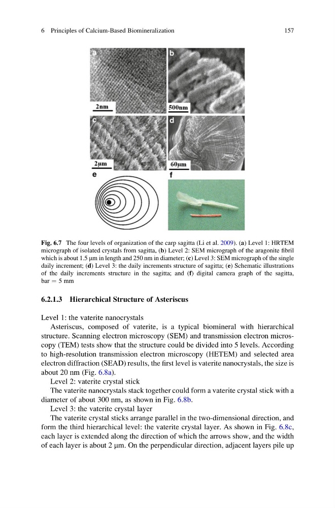
Fig. 6.7 The four levels of organization of the carp sagitta (Li et al. 2009). (a) Level 1: HRTEM
micrograph of isolated crystals from sagitta, (b) Level 2: SEM micrograph of the aragonite fibril
which is about 1.5 mm in length and 250 nm in diameter; (c) Level 3: SEM micrograph of the single
daily increment; (d) Level 3: the daily increments structure of sagitta; (e) Schematic illustrations
of the daily increments structure in the sagitta; and (f) digital camera graph of the sagitta,
bar ¼ 5 mm
6 Principles of Calcium-Based Biomineralization
157
Level 1: the vaterite nanocrystals
Asteriscus, composed of vaterite, is a typical biomineral with hierarchical
structure. Scanning electron microscopy (SEM) and transmission electron microscopy (TEM) tests show that the structure could be divided into 5 levels. According
to high-resolution transmission electron microscopy (HETEM) and selected area
electron diffraction (SEAD) results, the first level is vaterite nanocrystals, the size is
about 20 nm (Fig. 6.8a).
Level 2: vaterite crystal stick
The vaterite nanocrystals stack together could form a vaterite crystal stick with a
diameter of about 300 nm, as shown in Fig. 6.8b.
Level 3: the vaterite crystal layer
The vaterite crystal sticks arrange parallel in the two-dimensional direction, and
form the third hierarchical level: the vaterite crystal layer. As shown in Fig. 6.8c,
each layer is extended along the direction of which the arrows show, and the width
of each layer is about 2 mm. On the perpendicular direction, adjacent layers pile up
Fig. 6.7 The four levels of organization of the carp sagitta (Li et al. 2009). (a) Level 1: HRTEM
micrograph of isolated crystals from sagitta, (b) Level 2: SEM micrograph of the aragonite fibril
which is about 1.5 mm in length and 250 nm in diameter; (c) Level 3: SEM micrograph of the single
daily increment; (d) Level 3: the daily increments structure of sagitta; (e) Schematic illustrations
of the daily increments structure in the sagitta; and (f) digital camera graph of the sagitta,
bar ¼ 5 mm
6 Principles of Calcium-Based Biomineralization
157