

154
Horst Bleckmann et a!.
60
40
20
...-.
0
(/)
-
20
~
Q)
- Ctl
a: 10
Q)
0>
....
Ctl
..c
0
()
(/)
0 20
10
0
? __ __J
-4
-2
0
2
4
6
8
10
Sphere Location [em]
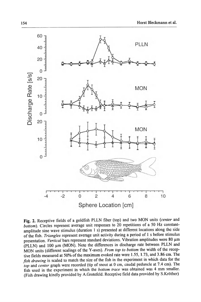
Fig. 2. Receptive fields of a goldfish PLLN fiber (top) and two MON units (center and
bottom). Circles represent average unit responses to 20 repetitions of a 50 Hz constantamplitude sine wave stimulus (duration I s) presented at different locations along the side
of the fish . Triangles represent average unit activity during a period of I s before stimulus
presentation. Vertical bars represent standard deviations. Vibration amplitudes were 80 11m
(PLLN) and I 00 11m (MON). Note the differences in discharge rate between PLLN and
MON units (different scalings of theY-axes). From top to bottom the width of the receptive fields measured at 50% of the maximum evoked rate were 1.55, 1.73, and 3.86 em. The
fish drawing is scaled to match the size of the fish in the experiment in which data for the
top and center graph were recorded (tip of snout at 0 em, caudal peduncle at 7.4 em). The
fish used in the experiment in which the bottom trace was obtained was 4 mm smaller.
(Fish drawing kindly provided by A.Grotefeld. Receptive field data provided by S.Krother)
Horst Bleckmann et a!.
60
40
20
...-.
0
(/)
-
20
~
Q)
- Ctl
a: 10
Q)
0>
....
Ctl
..c
0
()
(/)
0 20
10
0
? __ __J
-4
-2
0
2
4
6
8
10
Sphere Location [em]
Fig. 2. Receptive fields of a goldfish PLLN fiber (top) and two MON units (center and
bottom). Circles represent average unit responses to 20 repetitions of a 50 Hz constantamplitude sine wave stimulus (duration I s) presented at different locations along the side
of the fish . Triangles represent average unit activity during a period of I s before stimulus
presentation. Vertical bars represent standard deviations. Vibration amplitudes were 80 11m
(PLLN) and I 00 11m (MON). Note the differences in discharge rate between PLLN and
MON units (different scalings of theY-axes). From top to bottom the width of the receptive fields measured at 50% of the maximum evoked rate were 1.55, 1.73, and 3.86 em. The
fish drawing is scaled to match the size of the fish in the experiment in which data for the
top and center graph were recorded (tip of snout at 0 em, caudal peduncle at 7.4 em). The
fish used in the experiment in which the bottom trace was obtained was 4 mm smaller.
(Fish drawing kindly provided by A.Grotefeld. Receptive field data provided by S.Krother)