

Transgenic Technology in Marine Organisms 13.4 Biotechnological Application 401
Part B | 13.4
ful transgenic zebrafish displaying whole-body green,
red, yellow, or orange fluorescent colors in daylight,
dim light. or UV light [13.113, 114]. Some of these
fish have been sterilized to avoid contaminating the
wild population if they should accidentally be released
from aquaria to the environments [13.115]. For research purposes, mutant zebrafish and medaka with
less interfering pigmentation for optical observation
have also been developed. One such example is a line
of see-through transgenic medaka that are transparent
throughout their entire life, thus allowing clear visualization of GFP that is introduced into the fish as
a reporter [13.116].
While studies on enhancing body color variation in
ornamental fish were traditionally conducted in medaka
and zebrafish, very little has been accomplished in
mid-size or larger ornamental fish species. By coinjecting equal volumes of Tol2 transposase mRNA
(50 ng=L) into one-cell stage angelfish embryos with
a Tol2 transposon-mediated transgene construct containing a muscle-specific zebrafish CKMb 2:4 kb promoter/enhancer linked to Acropora coral red fluorescent
protein cDNA with SV40 polyA tail, we succeeded
in producing strains of pink body color transgenic angelfish expressing the Acropora coral red fluorescent
protein (Fig. 13.13). Furthermore, a strain of pink body
color transgenic lionhead fish, another strain of midsize ornamental fish, has also been produced by J.Y.
Chen and J.L. Wu in the Institute of Cellular and Organismal Biology, Academia Sinica, Taiwan (personal
communication).
13.4.4 Models for Studying Human Diseases
In order to provide a general proof of principle or
to reproduce specific aspects of human diseases, animal models have long been used to identify diseaseassociated factors or genes, or to screen new treatments
for these diseases [13.117–119]. In addition to primate and murine models, zebrafish have been used to
study human diseases for decades, particularly in cancer research and drug discovery [13.120, 121]. In recent
years, transgenic zebrafish are emerging as a valuable
model for studying human diseases, especially cardiac
and neurodegenerative diseases and cancers [13.122–
125]. In humans, tau proteins function to stabilize
microtubules in neurons of the central nervous system.
When tau proteins are defective, they result in dementias such as Alzheimer’s disease. However, the early
functional consequences of tau protein alterations in
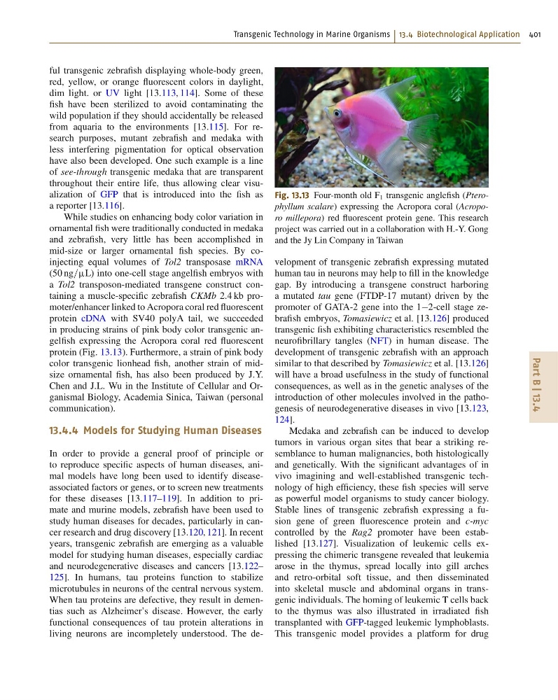
living neurons are incompletely understood. The deFig. 13.13 Four-month old F 1 transgenic anglefish (Pterophyllum scalare) expressing the Acropora coral (Acroporo millepora) red fluorescent protein gene. This research
project was carried out in a collaboration with H.-Y. Gong
and the Jy Lin Company in Taiwan
velopment of transgenic zebrafish expressing mutated
human tau in neurons may help to fill in the knowledge
gap. By introducing a transgene construct harboring
a mutated tau gene (FTDP-17 mutant) driven by the
promoter of GATA-2 gene into the 12-cell stage zebrafish embryos, Tomasiewicz et al. [13.126] produced
transgenic fish exhibiting characteristics resembled the
neurofibrillary tangles (NFT) in human disease. The
development of transgenic zebrafish with an approach
similar to that described by Tomasiewicz et al. [13.126]
will have a broad usefulness in the study of functional
consequences, as well as in the genetic analyses of the
introduction of other molecules involved in the pathogenesis of neurodegenerative diseases in vivo [13.123,
124].
Medaka and zebrafish can be induced to develop
tumors in various organ sites that bear a striking resemblance to human malignancies, both histologically
and genetically. With the significant advantages of in
vivo imagining and well-established transgenic technology of high efficiency, these fish species will serve
as powerful model organisms to study cancer biology.
Stable lines of transgenic zebrafish expressing a fusion gene of green fluorescence protein and c-myc
controlled by the Rag2 promoter have been established [13.127]. Visualization of leukemic cells expressing the chimeric transgene revealed that leukemia
arose in the thymus, spread locally into gill arches
and retro-orbital soft tissue, and then disseminated
into skeletal muscle and abdominal organs in transgenic individuals. The homing of leukemic T cells back
to the thymus was also illustrated in irradiated fish
transplanted with GFP-tagged leukemic lymphoblasts.
This transgenic model provides a platform for drug
Part B | 13.4
ful transgenic zebrafish displaying whole-body green,
red, yellow, or orange fluorescent colors in daylight,
dim light. or UV light [13.113, 114]. Some of these
fish have been sterilized to avoid contaminating the
wild population if they should accidentally be released
from aquaria to the environments [13.115]. For research purposes, mutant zebrafish and medaka with
less interfering pigmentation for optical observation
have also been developed. One such example is a line
of see-through transgenic medaka that are transparent
throughout their entire life, thus allowing clear visualization of GFP that is introduced into the fish as
a reporter [13.116].
While studies on enhancing body color variation in
ornamental fish were traditionally conducted in medaka
and zebrafish, very little has been accomplished in
mid-size or larger ornamental fish species. By coinjecting equal volumes of Tol2 transposase mRNA
(50 ng=L) into one-cell stage angelfish embryos with
a Tol2 transposon-mediated transgene construct containing a muscle-specific zebrafish CKMb 2:4 kb promoter/enhancer linked to Acropora coral red fluorescent
protein cDNA with SV40 polyA tail, we succeeded
in producing strains of pink body color transgenic angelfish expressing the Acropora coral red fluorescent
protein (Fig. 13.13). Furthermore, a strain of pink body
color transgenic lionhead fish, another strain of midsize ornamental fish, has also been produced by J.Y.
Chen and J.L. Wu in the Institute of Cellular and Organismal Biology, Academia Sinica, Taiwan (personal
communication).
13.4.4 Models for Studying Human Diseases
In order to provide a general proof of principle or
to reproduce specific aspects of human diseases, animal models have long been used to identify diseaseassociated factors or genes, or to screen new treatments
for these diseases [13.117–119]. In addition to primate and murine models, zebrafish have been used to
study human diseases for decades, particularly in cancer research and drug discovery [13.120, 121]. In recent
years, transgenic zebrafish are emerging as a valuable
model for studying human diseases, especially cardiac
and neurodegenerative diseases and cancers [13.122–
125]. In humans, tau proteins function to stabilize
microtubules in neurons of the central nervous system.
When tau proteins are defective, they result in dementias such as Alzheimer’s disease. However, the early
functional consequences of tau protein alterations in
living neurons are incompletely understood. The deFig. 13.13 Four-month old F 1 transgenic anglefish (Pterophyllum scalare) expressing the Acropora coral (Acroporo millepora) red fluorescent protein gene. This research
project was carried out in a collaboration with H.-Y. Gong
and the Jy Lin Company in Taiwan
velopment of transgenic zebrafish expressing mutated
human tau in neurons may help to fill in the knowledge
gap. By introducing a transgene construct harboring
a mutated tau gene (FTDP-17 mutant) driven by the
promoter of GATA-2 gene into the 12-cell stage zebrafish embryos, Tomasiewicz et al. [13.126] produced
transgenic fish exhibiting characteristics resembled the
neurofibrillary tangles (NFT) in human disease. The
development of transgenic zebrafish with an approach
similar to that described by Tomasiewicz et al. [13.126]
will have a broad usefulness in the study of functional
consequences, as well as in the genetic analyses of the
introduction of other molecules involved in the pathogenesis of neurodegenerative diseases in vivo [13.123,
124].
Medaka and zebrafish can be induced to develop
tumors in various organ sites that bear a striking resemblance to human malignancies, both histologically
and genetically. With the significant advantages of in
vivo imagining and well-established transgenic technology of high efficiency, these fish species will serve
as powerful model organisms to study cancer biology.
Stable lines of transgenic zebrafish expressing a fusion gene of green fluorescence protein and c-myc
controlled by the Rag2 promoter have been established [13.127]. Visualization of leukemic cells expressing the chimeric transgene revealed that leukemia
arose in the thymus, spread locally into gill arches
and retro-orbital soft tissue, and then disseminated
into skeletal muscle and abdominal organs in transgenic individuals. The homing of leukemic T cells back
to the thymus was also illustrated in irradiated fish
transplanted with GFP-tagged leukemic lymphoblasts.
This transgenic model provides a platform for drug