

149
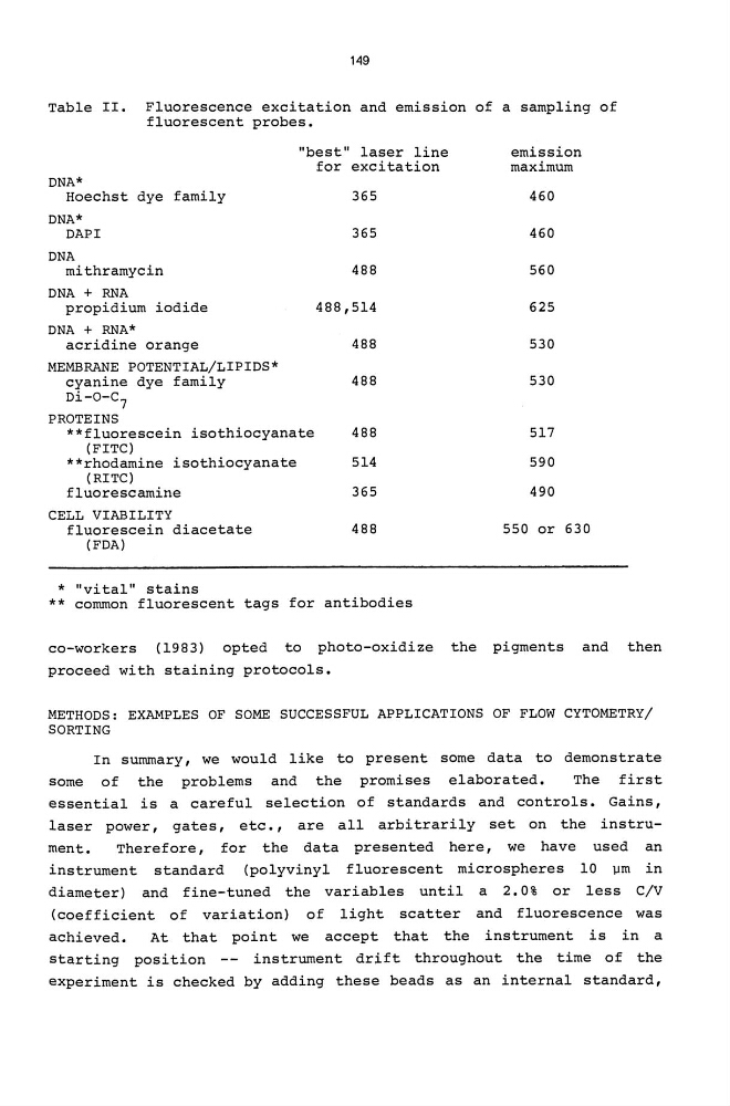
Table II. Fluorescence excitation and emission of a sampling of
fluorescent probes.
DNA*
"best" laser line
for excitation
Hoechst dye family
365
DNA*
DAPI
365
DNA
mithramycin
488
DNA + RNA
propidium iodide
488,514
DNA + RNA*
acridine orange
488
MEMBRANE POTENTIAL/LIPIDS*
cyanine dye family
488
Di-O-C 7
PROTEINS
**fluorescein isothiocyanate
488
(FITC)
**rhodamine isothiocyanate
514
(RITC)
fluorescamine
365
CELL VIABILITY
fluorescein diacetate
(FDA)
* "vital" stains
488
** common fluorescent tags for antibodies
emission
maximum
460
460
560
625
530
530
517
590
490
550 or 630
co-workers (1983) opted to photo-oxidize the pigments and then
proceed with staining protocols.
METHODS: EXAMPLES OF SOME SUCCESSFUL APPLICATIONS OF FLOW CYTOMETRY/
SORTING
In summary, we would like to present some data to demonstrate
some of the problems and the promises elaborated.
The first
essential is a careful selection of standards and controls. Gains,
laser power, gates, etc., are all arbitrarily set on the instrument.
Therefore, for the data presented here, we have used an
instrument standard (polyvinyl fluorescent microspheres 10 \lm in
diameter) and fine-tuned the variables until a 2.0% or less C/V
(coefficient of variation) of light scatter and fluorescence was
achieved.
At that point we accept that the instrument is in a
starting position -- instrument drift throughout the time of the
experiment is checked by adding these beads as an internal standard,
Table II. Fluorescence excitation and emission of a sampling of
fluorescent probes.
DNA*
"best" laser line
for excitation
Hoechst dye family
365
DNA*
DAPI
365
DNA
mithramycin
488
DNA + RNA
propidium iodide
488,514
DNA + RNA*
acridine orange
488
MEMBRANE POTENTIAL/LIPIDS*
cyanine dye family
488
Di-O-C 7
PROTEINS
**fluorescein isothiocyanate
488
(FITC)
**rhodamine isothiocyanate
514
(RITC)
fluorescamine
365
CELL VIABILITY
fluorescein diacetate
(FDA)
* "vital" stains
488
** common fluorescent tags for antibodies
emission
maximum
460
460
560
625
530
530
517
590
490
550 or 630
co-workers (1983) opted to photo-oxidize the pigments and then
proceed with staining protocols.
METHODS: EXAMPLES OF SOME SUCCESSFUL APPLICATIONS OF FLOW CYTOMETRY/
SORTING
In summary, we would like to present some data to demonstrate
some of the problems and the promises elaborated.
The first
essential is a careful selection of standards and controls. Gains,
laser power, gates, etc., are all arbitrarily set on the instrument.
Therefore, for the data presented here, we have used an
instrument standard (polyvinyl fluorescent microspheres 10 \lm in
diameter) and fine-tuned the variables until a 2.0% or less C/V
(coefficient of variation) of light scatter and fluorescence was
achieved.
At that point we accept that the instrument is in a
starting position -- instrument drift throughout the time of the
experiment is checked by adding these beads as an internal standard,