

130
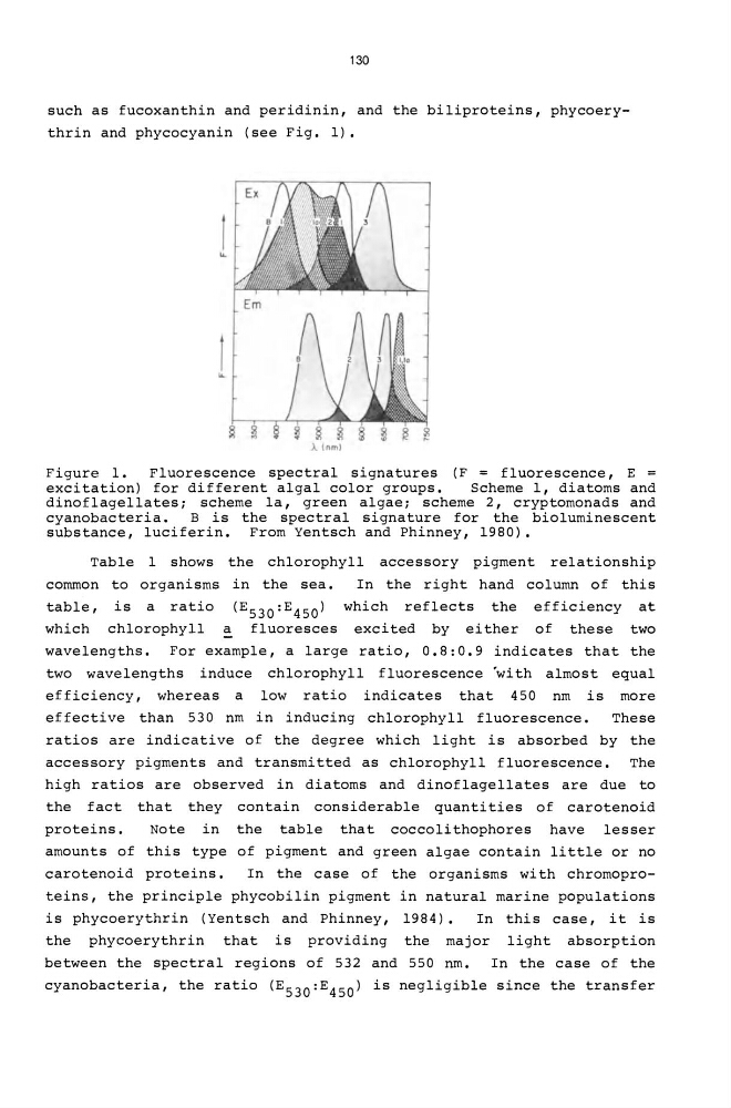
such as fucoxanthin and peridinin, and the biliproteins, phycoerythrin and phycocyanin (see Fig. 1) •
...
A (nm)
Figure 1.
Fluorescence spectral signatures (F = fluorescence, E =
excitation) for different algal color groups.
Scheme 1, diatoms and
dinoflagellates; scheme la, green algae; scheme 2, cryptomonads and
cyanobacteria.
B is the spectral signature for the bioluminescent
substance, luciferin. From Yentsch and Phinney, 1980).
Table 1 shows the chlorophyll accessory pigment relationship
common to organisms in the sea.
In the right hand column of this
table, is a ratio (E530:E450) which reflects the efficiency at
which chlorophyll ~ fluoresces excited by either of these two
wavelengths. For example, a large ratio, 0.8:0.9 indicates that the
two wavelengths induce chlorophyll fluorescence 'with almost equal
efficiency, whereas a low ratio indicates that 450 nm is more
effective than 530 nm in inducing chlorophyll fluorescence.
These
ratios are indicative of the degree which light is absorbed by the
accessory pigments and transmitted as chlorophyll fluorescence.
The
high ratios are observed in diatoms and dinoflagellates are due to
the fact that they contain considerable quanti ties of carotenoid
proteins.
Note in the table that coccolithophores have lesser
amounts of this type of pigment and green algae contain little or no
carotenoid proteins.
In the case of the organisms with chromoproteins, the principle phycobilin pigment in natural marine populations
is phycoerythrin (Yentsch and Phinney, 1984).
In this case, it is
the phycoerythrin that is providing the major light absorption
between the spectral regions of 532 and 550 nm.
In the case of the
cyanobacteria, the ratio (E530 : E450) is negligible since the transfer
such as fucoxanthin and peridinin, and the biliproteins, phycoerythrin and phycocyanin (see Fig. 1) •
...
A (nm)
Figure 1.
Fluorescence spectral signatures (F = fluorescence, E =
excitation) for different algal color groups.
Scheme 1, diatoms and
dinoflagellates; scheme la, green algae; scheme 2, cryptomonads and
cyanobacteria.
B is the spectral signature for the bioluminescent
substance, luciferin. From Yentsch and Phinney, 1980).
Table 1 shows the chlorophyll accessory pigment relationship
common to organisms in the sea.
In the right hand column of this
table, is a ratio (E530:E450) which reflects the efficiency at
which chlorophyll ~ fluoresces excited by either of these two
wavelengths. For example, a large ratio, 0.8:0.9 indicates that the
two wavelengths induce chlorophyll fluorescence 'with almost equal
efficiency, whereas a low ratio indicates that 450 nm is more
effective than 530 nm in inducing chlorophyll fluorescence.
These
ratios are indicative of the degree which light is absorbed by the
accessory pigments and transmitted as chlorophyll fluorescence.
The
high ratios are observed in diatoms and dinoflagellates are due to
the fact that they contain considerable quanti ties of carotenoid
proteins.
Note in the table that coccolithophores have lesser
amounts of this type of pigment and green algae contain little or no
carotenoid proteins.
In the case of the organisms with chromoproteins, the principle phycobilin pigment in natural marine populations
is phycoerythrin (Yentsch and Phinney, 1984).
In this case, it is
the phycoerythrin that is providing the major light absorption
between the spectral regions of 532 and 550 nm.
In the case of the
cyanobacteria, the ratio (E530 : E450) is negligible since the transfer