

202
The Indian work on the taxonomy, phenology
and distribution of water moulds has been initiated by Chaudhuri and Kochhar ( 1935 ) and followed by Chaudhuri and Lotus ( 1936 ), Hamid
( 1942 ), Dayal and Thakurji ( 1966 , 1968 ),
Srivastava ( 1967a ), Khulbe and Bhargava ( 1977 ),
Prabhuji and Srivastava ( 1977 , 1978 ), Khulbe
( 1980 ), Prabhuji ( 1984a , b ), Prabhuji et al.
( 1984 ), Prabhuji and Sinha ( 1993 , 1994 ) and
Prabhuji ( 2005 ). However, the studies made on
water moulds in India, particularly the members
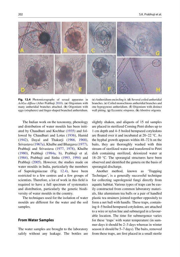
of Saprolegniaceae (Fig. 12.4 ), have been
restricted to a few centres and a few groups of
scientists. Therefore, a lot of work in this fi eld is
required to have a full spectrum of systematics
and distribution, particularly the genetic biodiversity of water moulds in the country.
The techniques used for the isolation of water
moulds are different for the water and the soil
samples.
From Water Samples
The water samples are brought to the laboratory
safely without any leakage. The bottles are
slightly shaken, and aliquots of 15 ml samples
are placed in sterilized Corning Petri dishes up to
1 cm depth and 4–5 boiled hempseed cotyledons
are fl oated over it and incubated at 20–22 °C. As
the hyphal growth appears within 48–72 h on the
baits, they are thoroughly washed with thin
stream of sterilized water and transferred to Petri
dish containing sterilized, deionized water at
18–20 °C. The sporangial structures have been
observed and identifi ed the genera on the basis of
sporangial discharge.
Another method, known as ‘Trapping
Technique’, is a generally successful technique
for collecting saprolegnoid fungi directly from
aquatic habitat. Various types of traps can be easily constructed from common laboratory materials, like aluminium tea balls or a pair of handled
plastic tea strainers jointed together oppositely to
form a net ball with handle. These traps, containing 4–5 boiled hempseed cotyledons, are attached
to a wire or nylon line and submerged in a favourable location. The time for submergence varies
for these ‘traps’ with water temperature (in summer days it should be 2–3 days whereas in winter
season it should be 5–7 days). The baits, removed
from these traps, are fi rst placed in a small sterile
Fig. 12.4 Photomicrographs of sexual apparatus in
Achlya diffusa (After Prabhuji 2010 ). ( a ) Oögonium with
many antheridial branches attached. ( b ) Oögonium with
eggs (oöspheres) and fi nger-shaped branched antheridium.
( c ) Antheridium encircling it. ( d ) Several coiled antheridial
branches. ( e ) Coiled monoclinous antheridial branches and
one hypogynous antheridium. ( f ) Oögonium with distinct
wall pitting. ( g ) Eccentric oöspores. ( h ) Abortive oögonia
S.K. Prabhuji et al.
The Indian work on the taxonomy, phenology
and distribution of water moulds has been initiated by Chaudhuri and Kochhar ( 1935 ) and followed by Chaudhuri and Lotus ( 1936 ), Hamid
( 1942 ), Dayal and Thakurji ( 1966 , 1968 ),
Srivastava ( 1967a ), Khulbe and Bhargava ( 1977 ),
Prabhuji and Srivastava ( 1977 , 1978 ), Khulbe
( 1980 ), Prabhuji ( 1984a , b ), Prabhuji et al.
( 1984 ), Prabhuji and Sinha ( 1993 , 1994 ) and
Prabhuji ( 2005 ). However, the studies made on
water moulds in India, particularly the members
of Saprolegniaceae (Fig. 12.4 ), have been
restricted to a few centres and a few groups of
scientists. Therefore, a lot of work in this fi eld is
required to have a full spectrum of systematics
and distribution, particularly the genetic biodiversity of water moulds in the country.
The techniques used for the isolation of water
moulds are different for the water and the soil
samples.
From Water Samples
The water samples are brought to the laboratory
safely without any leakage. The bottles are
slightly shaken, and aliquots of 15 ml samples
are placed in sterilized Corning Petri dishes up to
1 cm depth and 4–5 boiled hempseed cotyledons
are fl oated over it and incubated at 20–22 °C. As
the hyphal growth appears within 48–72 h on the
baits, they are thoroughly washed with thin
stream of sterilized water and transferred to Petri
dish containing sterilized, deionized water at
18–20 °C. The sporangial structures have been
observed and identifi ed the genera on the basis of
sporangial discharge.
Another method, known as ‘Trapping
Technique’, is a generally successful technique
for collecting saprolegnoid fungi directly from
aquatic habitat. Various types of traps can be easily constructed from common laboratory materials, like aluminium tea balls or a pair of handled
plastic tea strainers jointed together oppositely to
form a net ball with handle. These traps, containing 4–5 boiled hempseed cotyledons, are attached
to a wire or nylon line and submerged in a favourable location. The time for submergence varies
for these ‘traps’ with water temperature (in summer days it should be 2–3 days whereas in winter
season it should be 5–7 days). The baits, removed
from these traps, are fi rst placed in a small sterile
Fig. 12.4 Photomicrographs of sexual apparatus in
Achlya diffusa (After Prabhuji 2010 ). ( a ) Oögonium with
many antheridial branches attached. ( b ) Oögonium with
eggs (oöspheres) and fi nger-shaped branched antheridium.
( c ) Antheridium encircling it. ( d ) Several coiled antheridial
branches. ( e ) Coiled monoclinous antheridial branches and
one hypogynous antheridium. ( f ) Oögonium with distinct
wall pitting. ( g ) Eccentric oöspores. ( h ) Abortive oögonia
S.K. Prabhuji et al.