

50
U. Schreiber et al.
this purpose, see, e.g., Krause and Weis 1991; Schreiber and Bilger 1992).
Also, this chapter is not written for specialists in the field of fluorescence
research. Rather, it is intended to give an introduction to nonspecialists on
major aspects of the chlorophyll fluorescence indicator function, on the
methods developed for quenching analysis and on the physiologically relevant information obtained by fluorescence measurements.
3.2 Indicator Function of Chlorophyll Fluorescence
Contrary to chlorophyll in solution, chlorophyll in vivo displays large changes
in fluorescence yield upon illumination (Kautsky and Hirsch 1931). In vivo,
chlorophyll exists in the form of pigment protein complexes which are
embedded in the thylakoid membrane and funnel their excitation energy
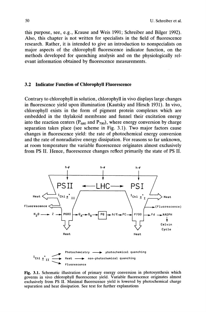
into the reaction centers (P680 and P700), where energy conversion by charge
separation takes place (see scheme in Fig. 3.1). Two major factors cause
changes in fluorescence yield: the rate of photochemical energy conversion
and the rate of nonradiative energy dissipation. For reasons so far unknown,
at room temperature the variable fluorescence originates almost exclusively
from PS II. Hence, fluorescence changes reflect primarily the state of PS II.
~LHC----"
Heat
Heat
Fluorescence
QA-QB~b/f __ PC
Heat
Heat
Photochemistry ~ photochemical quenching
Heat --.... non-photochemical quenching
Fluorescence
Fig. 3.1. Schematic illustration of primary energy conversion in photosynthesis which
governs in vivo chlorophyll fluorescence yield. Variable fluorescence originates almost
exclusively from PS II. Maximal fluorescence yield is lowered by photochemical charge
separation and heat dissipation. See text for further explanations
U. Schreiber et al.
this purpose, see, e.g., Krause and Weis 1991; Schreiber and Bilger 1992).
Also, this chapter is not written for specialists in the field of fluorescence
research. Rather, it is intended to give an introduction to nonspecialists on
major aspects of the chlorophyll fluorescence indicator function, on the
methods developed for quenching analysis and on the physiologically relevant information obtained by fluorescence measurements.
3.2 Indicator Function of Chlorophyll Fluorescence
Contrary to chlorophyll in solution, chlorophyll in vivo displays large changes
in fluorescence yield upon illumination (Kautsky and Hirsch 1931). In vivo,
chlorophyll exists in the form of pigment protein complexes which are
embedded in the thylakoid membrane and funnel their excitation energy
into the reaction centers (P680 and P700), where energy conversion by charge
separation takes place (see scheme in Fig. 3.1). Two major factors cause
changes in fluorescence yield: the rate of photochemical energy conversion
and the rate of nonradiative energy dissipation. For reasons so far unknown,
at room temperature the variable fluorescence originates almost exclusively
from PS II. Hence, fluorescence changes reflect primarily the state of PS II.
~LHC----"
Heat
Heat
Fluorescence
QA-QB~b/f __ PC
Heat
Heat
Photochemistry ~ photochemical quenching
Heat --.... non-photochemical quenching
Fluorescence
Fig. 3.1. Schematic illustration of primary energy conversion in photosynthesis which
governs in vivo chlorophyll fluorescence yield. Variable fluorescence originates almost
exclusively from PS II. Maximal fluorescence yield is lowered by photochemical charge
separation and heat dissipation. See text for further explanations