

3. Toxic Effects of Selenium in Fish
42
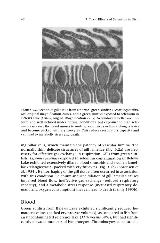
FIGURE 3.2. Section of gill tissue from a normal green sunfish (Lepomis cyanellus,
top, original magnification 240x), and a green sunfish exposed to selenium in
Belews Lake (bottom, original magnification 220x). Secondary lamellae are uniform and well defined under normal conditions, but exposure to high selenium can cause the blood sinuses to undergo extensive swelling (telangiectasia)
and become packed with erythrocytes. This reduces respiratory capacity and
can lead to metabolic stress and death.
ing pillar cells, which maintain the patency of vascular lumina. The
normally thin, delicate structures of gill lamellae (Fig. 3.2a) are necessary for effective gas exchange in respiration. Gills from green sunfish (Lepomis cyanellus) exposed to selenium contamination in Belews
Lake exhibited extensively dilated blood sinusoids and swollen lamellae (telangiectasia) packed with erythrocytes (Fig. 3.2b) (Sorensen et
al. 1984) . Hemorrhaging of the gill tissue often occurred in association
with this condition. Selenium-induced dilation of gill lamellae causes
impaired blood flow, ineffective gas exchange (reduced respiratory
capacity), and a metabolic stress response (increased respiratory demand and oxygen consumption) that can lead to death (Lemly 1993b).
Blood
Green sunfish from Belews Lake exhibited significantly reduced hematocrit values (packed erythrocyte volumes), as compared to fish from
an uncontaminated reference lake (33% versus 39%), but had significantly elevated numbers of lymphocytes. Thrombocytes constituted a
42
FIGURE 3.2. Section of gill tissue from a normal green sunfish (Lepomis cyanellus,
top, original magnification 240x), and a green sunfish exposed to selenium in
Belews Lake (bottom, original magnification 220x). Secondary lamellae are uniform and well defined under normal conditions, but exposure to high selenium can cause the blood sinuses to undergo extensive swelling (telangiectasia)
and become packed with erythrocytes. This reduces respiratory capacity and
can lead to metabolic stress and death.
ing pillar cells, which maintain the patency of vascular lumina. The
normally thin, delicate structures of gill lamellae (Fig. 3.2a) are necessary for effective gas exchange in respiration. Gills from green sunfish (Lepomis cyanellus) exposed to selenium contamination in Belews
Lake exhibited extensively dilated blood sinusoids and swollen lamellae (telangiectasia) packed with erythrocytes (Fig. 3.2b) (Sorensen et
al. 1984) . Hemorrhaging of the gill tissue often occurred in association
with this condition. Selenium-induced dilation of gill lamellae causes
impaired blood flow, ineffective gas exchange (reduced respiratory
capacity), and a metabolic stress response (increased respiratory demand and oxygen consumption) that can lead to death (Lemly 1993b).
Blood
Green sunfish from Belews Lake exhibited significantly reduced hematocrit values (packed erythrocyte volumes), as compared to fish from
an uncontaminated reference lake (33% versus 39%), but had significantly elevated numbers of lymphocytes. Thrombocytes constituted a