

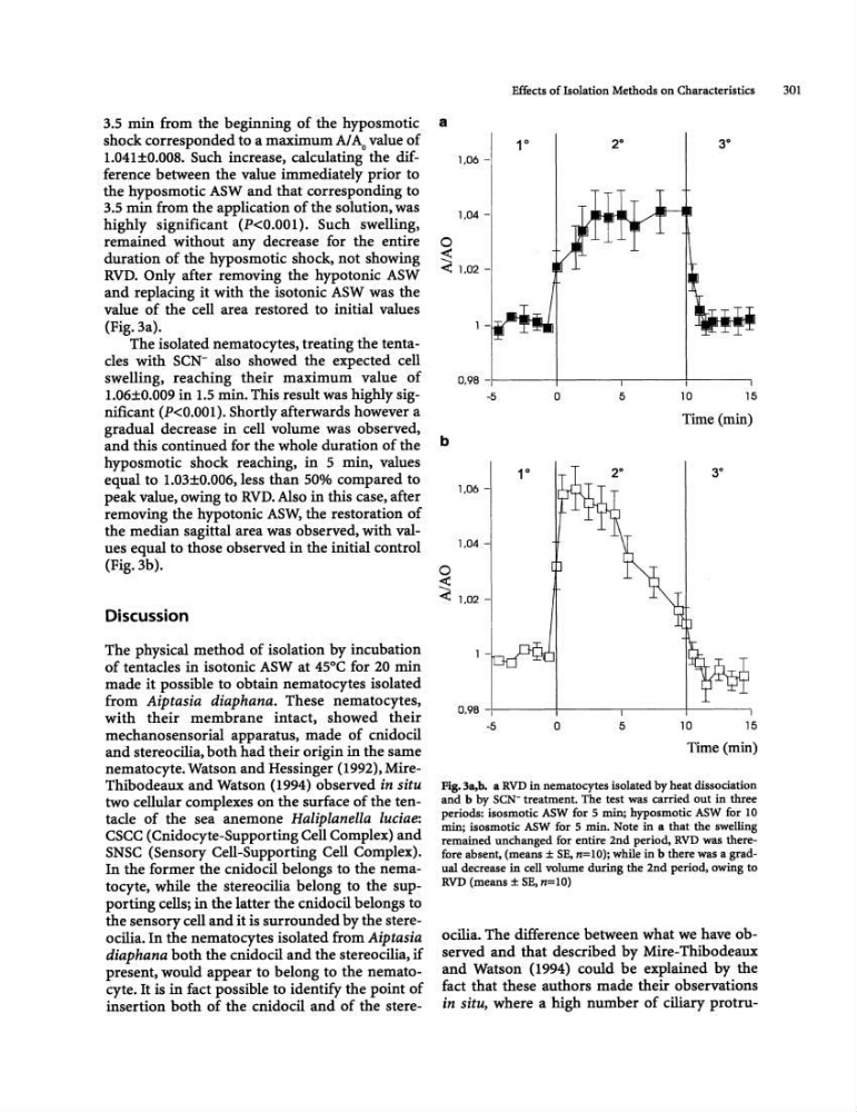
3.5 min from the beginning of the hyposmotic
shock corresponded to a maximum AI Ao value of
1.041±0.008. Such increase, calculating the difference between the value immediately prior to
the hyposmotic ASW and that corresponding to
3.5 min from the application of the solution, was
highly significant (P
duration of the hyposmotic shock, not showing
RVD. Only after removing the hypotonic ASW
and replacing it with the isotonic ASW was the
value of the cell area restored to initial values
(Fig.3a).
The isolated nematocytes, treating the tentacles with SCN- also showed the expected cell
swelling, reaching their maximum value of
1.06±O.009 in 1.5 min. This result was highly significant (P
and this continued for the whole duration of the
hyposmotic shock reaching, in 5 min, values
equal to 1.03±O.006, less than 50% compared to
peak value, owing to RVD. Also in this case, after
removing the hypotonic ASW, the restoration of
the median sagittal area was observed, with values equal to those observed in the initial control
(Fig.3b).
Discussion
The physical method of isolation by incubation
of tentacles in isotonic ASW at 45°C for 20 min
made it possible to obtain nematoeytes isolated
from Aiptasia diaphana. These nematocytes,
with their membrane intact, showed their
mechanosensorial apparatus. made of cnidocil
and stereocilia, both had their origin in the same
m:matocyte. Watson and Hessinger (1992), MireThibodeaux and Watson (1994) observed in situ
two cellular complexes on the surface of the tentacle of the sea anemone Haliplanella luciae:
CSCC (Cnidoeyte-Supporting Cell Complex) and
SNSC (Sensory Cell-Supporting Cell Complex).
In the former the cnidoci1 belongs to the nematocyte, while the stereocilia belong to the supporting cells; in the latter the cnidocil belongs to
the sensory cell and it is surrounded by the stereocilia. In the nematocytes isolated from Aiptasia
diaphana both the cnidocil and the stereocilia, if
present. would appear to belong to the nematoeyte. It is in fact possible to identify the point of
insertion both of the cnidocil and of the sterea
b
o
~
1,06 -
0,98 -I
-5
1.06 -
1,04 -
Effects of Isolation Methods on Characteristics
301
1"
3°
T
I
0
5
10
15
Time (min)
<: 1,02 -
1 -
0,98 -+1-----+-----,-------+----1
-5
0
5
10
15
Time (min)
Fig. la,b. a RVD in nematocytes isolated by heat dissociation
and b by SCN- treatment. The test was carried out in three
periods: isosmotic ASW fur 5 min; hyposmotic ASW for 10
min; isosmotic ASW for 5 min. Note in a that the swelling
remained unchanged for entire 2nd period. RVD was therefore absent, (means ± SE, n=lO); while in b there was a gradual decrease in cell volume during the 2nd period, owing to
RVD (means ± SE, n=10)
ocilia. The difference between what we have observed and that described by Mire-Thibodeaux
and Watson (1994) could be explained by the
fact that these authors made their observations
in situ, where a high number of ciliary protru-
shock corresponded to a maximum AI Ao value of
1.041±0.008. Such increase, calculating the difference between the value immediately prior to
the hyposmotic ASW and that corresponding to
3.5 min from the application of the solution, was
highly significant (P
duration of the hyposmotic shock, not showing
RVD. Only after removing the hypotonic ASW
and replacing it with the isotonic ASW was the
value of the cell area restored to initial values
(Fig.3a).
The isolated nematocytes, treating the tentacles with SCN- also showed the expected cell
swelling, reaching their maximum value of
1.06±O.009 in 1.5 min. This result was highly significant (P
and this continued for the whole duration of the
hyposmotic shock reaching, in 5 min, values
equal to 1.03±O.006, less than 50% compared to
peak value, owing to RVD. Also in this case, after
removing the hypotonic ASW, the restoration of
the median sagittal area was observed, with values equal to those observed in the initial control
(Fig.3b).
Discussion
The physical method of isolation by incubation
of tentacles in isotonic ASW at 45°C for 20 min
made it possible to obtain nematoeytes isolated
from Aiptasia diaphana. These nematocytes,
with their membrane intact, showed their
mechanosensorial apparatus. made of cnidocil
and stereocilia, both had their origin in the same
m:matocyte. Watson and Hessinger (1992), MireThibodeaux and Watson (1994) observed in situ
two cellular complexes on the surface of the tentacle of the sea anemone Haliplanella luciae:
CSCC (Cnidoeyte-Supporting Cell Complex) and
SNSC (Sensory Cell-Supporting Cell Complex).
In the former the cnidoci1 belongs to the nematocyte, while the stereocilia belong to the supporting cells; in the latter the cnidocil belongs to
the sensory cell and it is surrounded by the stereocilia. In the nematocytes isolated from Aiptasia
diaphana both the cnidocil and the stereocilia, if
present. would appear to belong to the nematoeyte. It is in fact possible to identify the point of
insertion both of the cnidocil and of the sterea
b
o
~
1,06 -
0,98 -I
-5
1.06 -
1,04 -
Effects of Isolation Methods on Characteristics
301
1"
3°
T
I
0
5
10
15
Time (min)
<: 1,02 -
1 -
0,98 -+1-----+-----,-------+----1
-5
0
5
10
15
Time (min)
Fig. la,b. a RVD in nematocytes isolated by heat dissociation
and b by SCN- treatment. The test was carried out in three
periods: isosmotic ASW fur 5 min; hyposmotic ASW for 10
min; isosmotic ASW for 5 min. Note in a that the swelling
remained unchanged for entire 2nd period. RVD was therefore absent, (means ± SE, n=lO); while in b there was a gradual decrease in cell volume during the 2nd period, owing to
RVD (means ± SE, n=10)
ocilia. The difference between what we have observed and that described by Mire-Thibodeaux
and Watson (1994) could be explained by the
fact that these authors made their observations
in situ, where a high number of ciliary protru-