

210
and distinguishing between fl uorescent compounds and thereby increasing the contrast
between target organisms.
4
Fluorescent Observation
4.1
Laboratory Test Using
a UV-LED Light
A UV-LED illumination and camera system have
been developed for taking fl uorescent photographs of deep-sea organisms. The wavelength of
the illumination light is 375 nm with a half-width
of 10 nm, and the photographs were obtained
using an ordinary compact digital camera. Deepsea organisms kept in an aquarium were used as
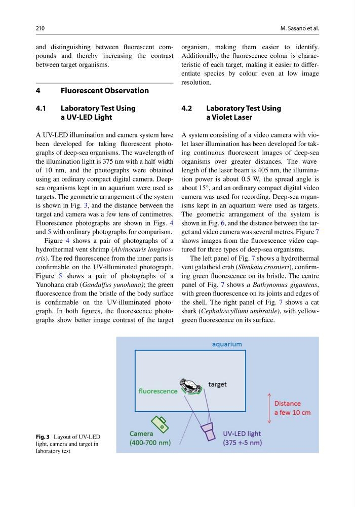
targets. The geometric arrangement of the system
is shown in Fig. 3 , and the distance between the
target and camera was a few tens of centimetres.
Fluorescence photographs are shown in Figs. 4
and 5 with ordinary photographs for comparison.
Figure 4 shows a pair of photographs of a
hydrothermal vent shrimp ( Alvinocaris longirostris ). The red fl uorescence from the inner parts is
confi rmable on the UV-illuminated photograph.
Figure 5 shows a pair of photographs of a
Yunohana crab ( Gandalfus yunohana) ; the green
fl uorescence from the bristle of the body surface
is confi rmable on the UV-illuminated photograph. In both fi gures, the fl uorescence photographs show better image contrast of the target
organism, making them easier to identify.
Additionally, the fl uorescence colour is characteristic of each target, making it easier to differentiate species by colour even at low image
resolution.
4.2
Laboratory Test Using
a Violet Laser
A system consisting of a video camera with violet laser illumination has been developed for taking continuous fl uorescent images of deep-sea
organisms over greater distances. The wavelength of the laser beam is 405 nm, the illumination power is about 0.5 W, the spread angle is
about 15°, and an ordinary compact digital video
camera was used for recording. Deep-sea organisms kept in an aquarium were used as targets.
The geometric arrangement of the system is
shown in Fig. 6 , and the distance between the target and video camera was several metres. Figure 7
shows images from the fl uorescence video captured for three types of deep-sea organisms.
The left panel of Fig. 7 shows a hydrothermal
vent galatheid crab ( Shinkaia crosnieri ), confi rming green fl uorescence on its bristle. The centre
panel of Fig. 7 shows a Bathynomus giganteus ,
with green fl uorescence on its joints and edges of
the shell. The right panel of Fig. 7 shows a cat
shark ( Cephaloscyllium umbratile) , with yellowgreen fl uorescence on its surface.
Fig. 3 Layout of UV-LED
light, camera and target in
laboratory test
M. Sasano et al.
and distinguishing between fl uorescent compounds and thereby increasing the contrast
between target organisms.
4
Fluorescent Observation
4.1
Laboratory Test Using
a UV-LED Light
A UV-LED illumination and camera system have
been developed for taking fl uorescent photographs of deep-sea organisms. The wavelength of
the illumination light is 375 nm with a half-width
of 10 nm, and the photographs were obtained
using an ordinary compact digital camera. Deepsea organisms kept in an aquarium were used as
targets. The geometric arrangement of the system
is shown in Fig. 3 , and the distance between the
target and camera was a few tens of centimetres.
Fluorescence photographs are shown in Figs. 4
and 5 with ordinary photographs for comparison.
Figure 4 shows a pair of photographs of a
hydrothermal vent shrimp ( Alvinocaris longirostris ). The red fl uorescence from the inner parts is
confi rmable on the UV-illuminated photograph.
Figure 5 shows a pair of photographs of a
Yunohana crab ( Gandalfus yunohana) ; the green
fl uorescence from the bristle of the body surface
is confi rmable on the UV-illuminated photograph. In both fi gures, the fl uorescence photographs show better image contrast of the target
organism, making them easier to identify.
Additionally, the fl uorescence colour is characteristic of each target, making it easier to differentiate species by colour even at low image
resolution.
4.2
Laboratory Test Using
a Violet Laser
A system consisting of a video camera with violet laser illumination has been developed for taking continuous fl uorescent images of deep-sea
organisms over greater distances. The wavelength of the laser beam is 405 nm, the illumination power is about 0.5 W, the spread angle is
about 15°, and an ordinary compact digital video
camera was used for recording. Deep-sea organisms kept in an aquarium were used as targets.
The geometric arrangement of the system is
shown in Fig. 6 , and the distance between the target and video camera was several metres. Figure 7
shows images from the fl uorescence video captured for three types of deep-sea organisms.
The left panel of Fig. 7 shows a hydrothermal
vent galatheid crab ( Shinkaia crosnieri ), confi rming green fl uorescence on its bristle. The centre
panel of Fig. 7 shows a Bathynomus giganteus ,
with green fl uorescence on its joints and edges of
the shell. The right panel of Fig. 7 shows a cat
shark ( Cephaloscyllium umbratile) , with yellowgreen fl uorescence on its surface.
Fig. 3 Layout of UV-LED
light, camera and target in
laboratory test
M. Sasano et al.