

The Heterotrimeric G Protein Genes of Caenorhabditis e/egans
19
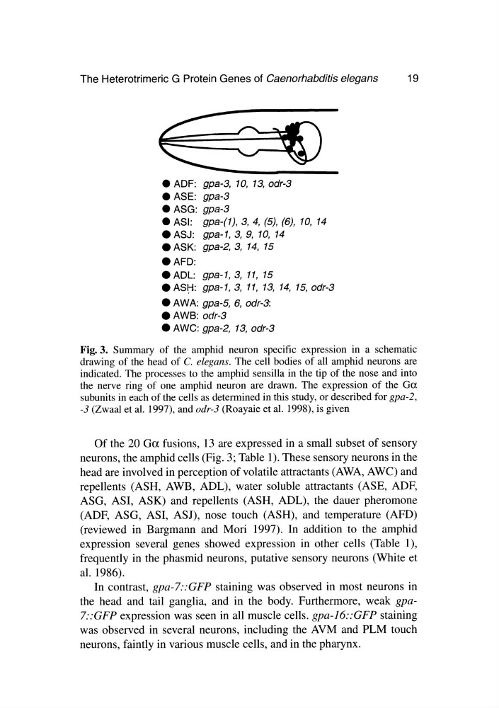
• ADF: gpa-3, 10, 13,odr-3
• ASE: gpa-3
• ASG: gpa-3
• ASI: gpa-(I), 3, 4, (5), (6), 10, 14
• ASJ: gpa-I,3, 9, 10, 14
• ASK: gpa-2,3, 14, 15
.AFD:
• ADL: gpa-I,3, 11, 15
• ASrt: gpa-I,3, 11, 13, 14, 15,odr-3
• AWA: gpa-5, 6, odr-3:
• AWB: odr-3
• AWe: gpa-2, 13,odr-3
Fig. 3. Summary of the amphid neuron specific expression in a schematic
drawing of the head of C. elegans. The cell bodies of all amphid neurons are
indicated. The processes to the amphid sensilla in the tip of the nose and into
the nerve ring of one amphid neuron are drawn. The expression of the Ga
subunits in each of the cells as determined in this study, or described for gpa-2,
-3 (Zwaal et al. 1997), and odr-3 (Roayaie et al. 1998), is given
Of the 20 Ga fusions, 13 are expressed in a small subset of sensory
neurons, the amphid cells (Fig. 3; Table 1). These sensory neurons in the
head are involved in perception of volatile attractants (AWA, AWe) and
repellents (ASH, AWB, ADL), water soluble attractants (ASE, ADF,
ASG, ASI, ASK) and repellents (ASH, ADL), the dauer pheromone
(ADF, ASG, AS I, ASJ), nose touch (ASH), and temperature (AFD)
(reviewed in Bargmann and Mori 1997). In addition to the amphid
expression several genes showed expression in other cells (Table 1),
frequently in the phasmid neurons, putative sensory neurons (White et
al. 1986).
In contrast, gpa-7::GFP staining was observed in most neurons in
the head and tail ganglia, and in the body. Furthermore, weak gpa7::GFP expression was seen in all muscle cells. gpa-16::GFP staining
was observed in several neurons, including the AVM and PLM touch
neurons, faintly in various muscle cells, and in the pharynx.
19
• ADF: gpa-3, 10, 13,odr-3
• ASE: gpa-3
• ASG: gpa-3
• ASI: gpa-(I), 3, 4, (5), (6), 10, 14
• ASJ: gpa-I,3, 9, 10, 14
• ASK: gpa-2,3, 14, 15
.AFD:
• ADL: gpa-I,3, 11, 15
• ASrt: gpa-I,3, 11, 13, 14, 15,odr-3
• AWA: gpa-5, 6, odr-3:
• AWB: odr-3
• AWe: gpa-2, 13,odr-3
Fig. 3. Summary of the amphid neuron specific expression in a schematic
drawing of the head of C. elegans. The cell bodies of all amphid neurons are
indicated. The processes to the amphid sensilla in the tip of the nose and into
the nerve ring of one amphid neuron are drawn. The expression of the Ga
subunits in each of the cells as determined in this study, or described for gpa-2,
-3 (Zwaal et al. 1997), and odr-3 (Roayaie et al. 1998), is given
Of the 20 Ga fusions, 13 are expressed in a small subset of sensory
neurons, the amphid cells (Fig. 3; Table 1). These sensory neurons in the
head are involved in perception of volatile attractants (AWA, AWe) and
repellents (ASH, AWB, ADL), water soluble attractants (ASE, ADF,
ASG, ASI, ASK) and repellents (ASH, ADL), the dauer pheromone
(ADF, ASG, AS I, ASJ), nose touch (ASH), and temperature (AFD)
(reviewed in Bargmann and Mori 1997). In addition to the amphid
expression several genes showed expression in other cells (Table 1),
frequently in the phasmid neurons, putative sensory neurons (White et
al. 1986).
In contrast, gpa-7::GFP staining was observed in most neurons in
the head and tail ganglia, and in the body. Furthermore, weak gpa7::GFP expression was seen in all muscle cells. gpa-16::GFP staining
was observed in several neurons, including the AVM and PLM touch
neurons, faintly in various muscle cells, and in the pharynx.