

166
A
B
dor al
ventral
Xenopus
chordin
Drosophila
dpp
C. Niehrs et al.
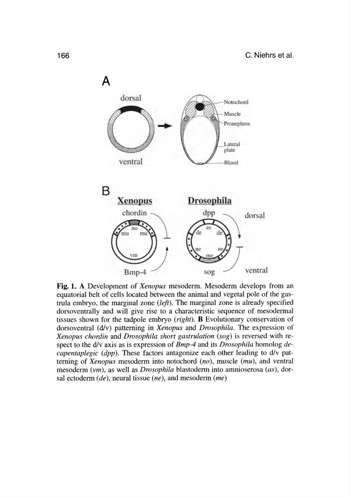
Fig. 1. A Development of Xenopus mesoderm. Mesoderm develops from an
equatorial belt of cells located between the animal and vegetal pole of the gastrula embryo, the marginal zone (left). The marginal zone is already specified
dorsoventrally and will give rise to a characteristic sequence of mesodermal
tissues shown for the tadpole embryo (right). B Evolutionary conservation of
dorsoventral (d/v) patterning in Xenopus and Drosophila. The expression of
Xenopus chordin and Drosophila short gastrulation (sog) is reversed with respect to the d/v axis as is expression of Bmp-4 and its Drosophila homolog decapentap/egic (dpp). These factors antagonize each other leading to d/v patterning of Xenopus mesoderm into notochord (no), muscle (mu), and ventral
mesoderm (vm), as well as Drosophila blastoderm into amnioserosa (as) , dorsal ectoderm (de) , neural tissue (ne) , and mesoderm (me)
A
B
dor al
ventral
Xenopus
chordin
Drosophila
dpp
C. Niehrs et al.
Fig. 1. A Development of Xenopus mesoderm. Mesoderm develops from an
equatorial belt of cells located between the animal and vegetal pole of the gastrula embryo, the marginal zone (left). The marginal zone is already specified
dorsoventrally and will give rise to a characteristic sequence of mesodermal
tissues shown for the tadpole embryo (right). B Evolutionary conservation of
dorsoventral (d/v) patterning in Xenopus and Drosophila. The expression of
Xenopus chordin and Drosophila short gastrulation (sog) is reversed with respect to the d/v axis as is expression of Bmp-4 and its Drosophila homolog decapentap/egic (dpp). These factors antagonize each other leading to d/v patterning of Xenopus mesoderm into notochord (no), muscle (mu), and ventral
mesoderm (vm), as well as Drosophila blastoderm into amnioserosa (as) , dorsal ectoderm (de) , neural tissue (ne) , and mesoderm (me)