

Genetic Dissection of Heart Development
113
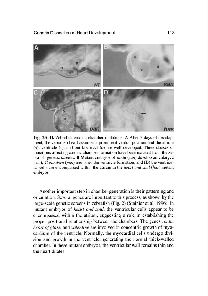
Fig. 2A-D. Zebrafish cardiac chamber mutations. A After 3 days of development, the zebrafish heart assumes a prominent ventral position and the atrium
(a) , ventricle (v), and outflow tract (0) are well developed. Three classes of
mutations affecting cardiac chamber formation have been isolated from the zebrafish genetic screens. B Mutant embryos of santa (san) develop an enlarged
heart. C pandora (pan) abolishes the ventricle formation, and (D) the ventricular cells are encompassed within the atrium in the heart and soul (has) mutant
embryos
Another important step in chamber generation is their patterning and
orientation. Several genes are important to this process, as shown by the
large-scale genetic screens in zebrafish (Fig. 2) (Stainier et al. 1996). In
mutant embryos of heart and soul, the ventricular cells appear to be
encompassed within the atrium, suggesting a role in establishing the
proper positional relationship between the chambers. The genes santa,
heart of glass, and valentine are involved in concentric growth of myocardium of the ventricle. Normally, the myocardial cells undergo division and growth in the ventricle, generating the normal thick-walled
chamber. In these mutant embryos, the ventricular wall remains thin and
the heart dilates.
113
Fig. 2A-D. Zebrafish cardiac chamber mutations. A After 3 days of development, the zebrafish heart assumes a prominent ventral position and the atrium
(a) , ventricle (v), and outflow tract (0) are well developed. Three classes of
mutations affecting cardiac chamber formation have been isolated from the zebrafish genetic screens. B Mutant embryos of santa (san) develop an enlarged
heart. C pandora (pan) abolishes the ventricle formation, and (D) the ventricular cells are encompassed within the atrium in the heart and soul (has) mutant
embryos
Another important step in chamber generation is their patterning and
orientation. Several genes are important to this process, as shown by the
large-scale genetic screens in zebrafish (Fig. 2) (Stainier et al. 1996). In
mutant embryos of heart and soul, the ventricular cells appear to be
encompassed within the atrium, suggesting a role in establishing the
proper positional relationship between the chambers. The genes santa,
heart of glass, and valentine are involved in concentric growth of myocardium of the ventricle. Normally, the myocardial cells undergo division and growth in the ventricle, generating the normal thick-walled
chamber. In these mutant embryos, the ventricular wall remains thin and
the heart dilates.