

110
J.-N. Chen and M. C. Fishman
A
B
,
/I'
' "
A
145
205
C
0
L
48hpf
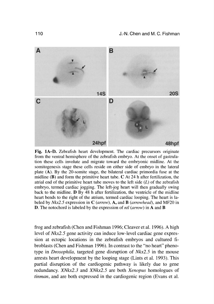
Fig. lA-D. Zebrafish heart development. The cardiac precursors ongmate
from the ventral hemisphere of the zebrafish embryo. At the onset of gastrulation these cells involute and migrate toward the embryonic midline. At the
somitogenesis stage these cells reside on either side of embryo in the lateral
plate (A). By the 20-somite stage, the bilateral cardiac primordia fuse at the
midline (B) and form the primitive heart tube. C At 24 h after fertilization, the
atrial end of the primitive heart tube moves to the left side (L) of the zebrafish
embryo, termed cardiac jogging. The left-jog heart will then gradually swing
back to the midline. D By 48 h after fertilization, the ventricle of the midline
heart bends to the right of the atrium, termed cardiac looping. The heart is labeled by Nkx2.5 expression in C (arrow), A, and B (arrowhead), and MF20 in
D. The notochord is labeled by the expression of nt[ (arrow) in A and B
frog and zebrafish (Chen and Fishman 1996; Cleaver et al. 1996). A high
level of Nkx2.5 gene activity can induce low-level cardiac gene expression at ectopic locations in the zebrafish embryos and cultured fibroblasts (Chen and Fishman 1996). In contrast to the "no heart" phenotype in Drosophila, targeted gene disruption of Nkx2.5 in the mouse
arrests heart development by the looping stage (Lints et al. 1993). This
partial disruption of the cardiogenic pathway is likely due to gene
redundancy. XNkx2.3 and XNkx2.5 are both Xenopus homologues of
tinman, and are both expressed in the cardiogenic region (Evans et al.
J.-N. Chen and M. C. Fishman
A
B
,
/I'
' "
A
145
205
C
0
L
48hpf
Fig. lA-D. Zebrafish heart development. The cardiac precursors ongmate
from the ventral hemisphere of the zebrafish embryo. At the onset of gastrulation these cells involute and migrate toward the embryonic midline. At the
somitogenesis stage these cells reside on either side of embryo in the lateral
plate (A). By the 20-somite stage, the bilateral cardiac primordia fuse at the
midline (B) and form the primitive heart tube. C At 24 h after fertilization, the
atrial end of the primitive heart tube moves to the left side (L) of the zebrafish
embryo, termed cardiac jogging. The left-jog heart will then gradually swing
back to the midline. D By 48 h after fertilization, the ventricle of the midline
heart bends to the right of the atrium, termed cardiac looping. The heart is labeled by Nkx2.5 expression in C (arrow), A, and B (arrowhead), and MF20 in
D. The notochord is labeled by the expression of nt[ (arrow) in A and B
frog and zebrafish (Chen and Fishman 1996; Cleaver et al. 1996). A high
level of Nkx2.5 gene activity can induce low-level cardiac gene expression at ectopic locations in the zebrafish embryos and cultured fibroblasts (Chen and Fishman 1996). In contrast to the "no heart" phenotype in Drosophila, targeted gene disruption of Nkx2.5 in the mouse
arrests heart development by the looping stage (Lints et al. 1993). This
partial disruption of the cardiogenic pathway is likely due to gene
redundancy. XNkx2.3 and XNkx2.5 are both Xenopus homologues of
tinman, and are both expressed in the cardiogenic region (Evans et al.