

238
Mohamed A1-Rubeai
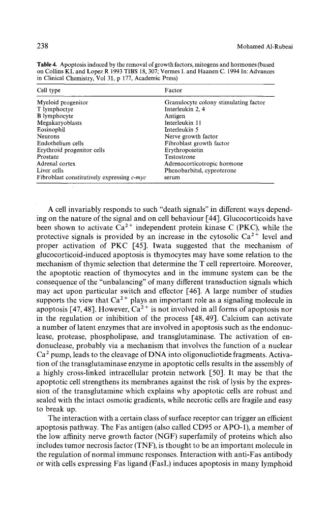
Table 4. Apoptosis induced by the removal of growth factors, mitogens and hormones (based
on Collins KL and Lopez R 1993 TIBS 18, 307; Vermes I. and Haanen C. 1994 In: Advances
in Clinical Chemistry, Vol 31, p 177, Academic Press)
Cell type
Factor
Myeloid progenitor
T lymphoctye
B lymphocyte
Megakaryoblasts
Eosinophil
Neurons
Endothelium cells
Erythroid progenitor cells
Prostate
Adrenal cortex
Liver cells
Fibroblast constitutively expressing c-myc
Granulocyte colony stimulating factor
Interleukin 2, 4
Antigen
Interleukin 11
Interleukin 5
Nerve growth factor
Fibroblast growth factor
Erythropoietin
Testostrone
Adrenocorticotropic hormone
Phenobarbital, cyproterone
serum
A cell invariably responds to such "death signals" in different ways depending on the nature of the signal and on cell behaviour [44]. Glucocorticoids have
been shown to activate Ca 2 + independent protein kinase C (PKC), while the
protective signals is provided by an increase in the cytosolic Ca 2 + level and
proper activation of PKC [45]. Iwata suggested that the mechanism of
glucocorticoid-induced apoptosis is thymocytes may have some relation to the
mechanism of thymic selection that determine the T cell reprertoire. Moreover,
the apoptotic reaction of thymocytes and in the immune system can be the
consequence of the "unbalancing" of many different transduction signals which
may act upon particular switch and effector [46]. A large number of studies
supports the view that Ca 2 + plays an important role as a signaling molecule in
apoptosis [47, 48]. However, Ca 2 + is not involved in all forms of apoptosis nor
in the regulation or inhibition of the process [48, 49]. Calcium can activate
a number of latent enzymes that are involved in apoptosis such as the endonuclease, protease, phospholipase, and transglutaminase. The activation of endonuclease, probably via a mechanism that involves the function of a nuclear
Ca 2 pump, leads to the cleavage of DNA into oligonucliotide fragments. Activation of the transglutaminase enzyme in apoptotic ceils results in the assembly of
a highly cross-linked intracellular protein network [50]. It may be that the
apoptotic cell strengthens its membranes against the risk of lysis by the expression of the transglutamine which explains why apoptotic cells are robust and
sealed with the intact osmotic gradients, while necrotic cells are fragile and easy
to break up.
The interaction with a certain class of surface receptor can trigger an efficient
apoptosis pathway. The Fas antigen (also called CD95 or APO-1), a member of
the low affinity nerve growth factor (NGF) superfamily of proteins which also
includes tumor necrosis factor (TNF), is thought to be an important molecule in
the regulation of normal immune responses. Interaction with anti-Fas antibody
or with cells expressing Fas ligand (FasL) induces apoptosis in many lymphoid
Mohamed A1-Rubeai
Table 4. Apoptosis induced by the removal of growth factors, mitogens and hormones (based
on Collins KL and Lopez R 1993 TIBS 18, 307; Vermes I. and Haanen C. 1994 In: Advances
in Clinical Chemistry, Vol 31, p 177, Academic Press)
Cell type
Factor
Myeloid progenitor
T lymphoctye
B lymphocyte
Megakaryoblasts
Eosinophil
Neurons
Endothelium cells
Erythroid progenitor cells
Prostate
Adrenal cortex
Liver cells
Fibroblast constitutively expressing c-myc
Granulocyte colony stimulating factor
Interleukin 2, 4
Antigen
Interleukin 11
Interleukin 5
Nerve growth factor
Fibroblast growth factor
Erythropoietin
Testostrone
Adrenocorticotropic hormone
Phenobarbital, cyproterone
serum
A cell invariably responds to such "death signals" in different ways depending on the nature of the signal and on cell behaviour [44]. Glucocorticoids have
been shown to activate Ca 2 + independent protein kinase C (PKC), while the
protective signals is provided by an increase in the cytosolic Ca 2 + level and
proper activation of PKC [45]. Iwata suggested that the mechanism of
glucocorticoid-induced apoptosis is thymocytes may have some relation to the
mechanism of thymic selection that determine the T cell reprertoire. Moreover,
the apoptotic reaction of thymocytes and in the immune system can be the
consequence of the "unbalancing" of many different transduction signals which
may act upon particular switch and effector [46]. A large number of studies
supports the view that Ca 2 + plays an important role as a signaling molecule in
apoptosis [47, 48]. However, Ca 2 + is not involved in all forms of apoptosis nor
in the regulation or inhibition of the process [48, 49]. Calcium can activate
a number of latent enzymes that are involved in apoptosis such as the endonuclease, protease, phospholipase, and transglutaminase. The activation of endonuclease, probably via a mechanism that involves the function of a nuclear
Ca 2 pump, leads to the cleavage of DNA into oligonucliotide fragments. Activation of the transglutaminase enzyme in apoptotic ceils results in the assembly of
a highly cross-linked intracellular protein network [50]. It may be that the
apoptotic cell strengthens its membranes against the risk of lysis by the expression of the transglutamine which explains why apoptotic cells are robust and
sealed with the intact osmotic gradients, while necrotic cells are fragile and easy
to break up.
The interaction with a certain class of surface receptor can trigger an efficient
apoptosis pathway. The Fas antigen (also called CD95 or APO-1), a member of
the low affinity nerve growth factor (NGF) superfamily of proteins which also
includes tumor necrosis factor (TNF), is thought to be an important molecule in
the regulation of normal immune responses. Interaction with anti-Fas antibody
or with cells expressing Fas ligand (FasL) induces apoptosis in many lymphoid