

76
700
600
c soo
8
~ 400
'"
~ 300
.:
200
100
600
400
600
.00
200
A
B
c
yl
200
y7
y4
y6
y3
\
000
y6
y4
y2
I
600
M. WILM et al.
b l 4
ylo
blS
x 10
yl4
y8
y9
b l 7
b lO
1000
ISoo
2000
miz, amu
D
bll
b12
80
b14
60
blS
y7
E
y8
'0
biZ
yl2
bl4
40
blS
30
20
800
ml1:, amu
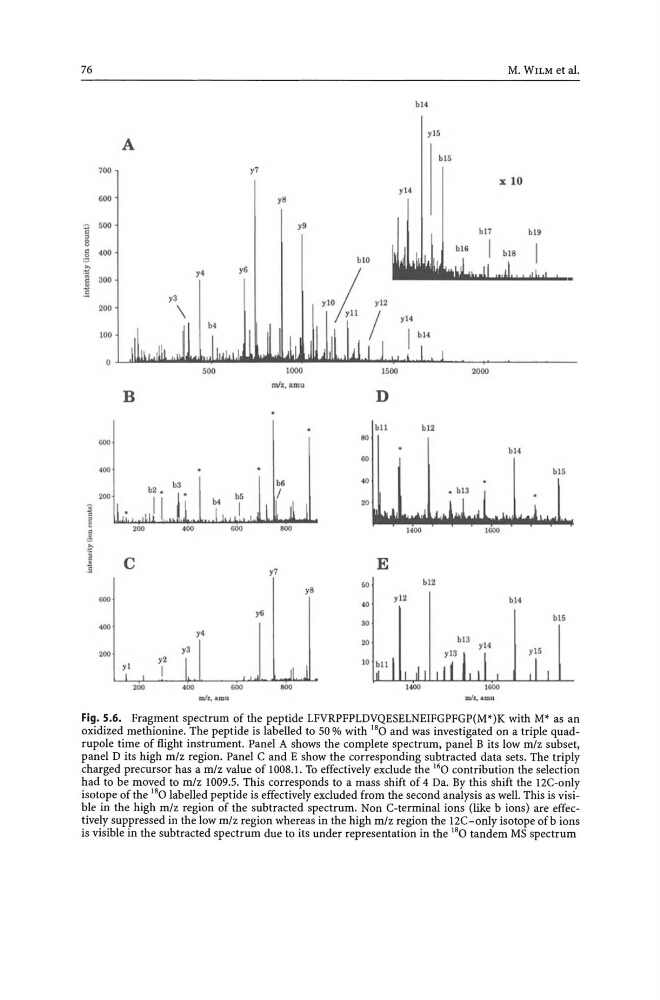
Fig. 5.6. Fragment spectrum of the peptide LFVRPFPLDVQESELNEIFGPFGP(M*)K with M* as an
oxidized methionine. The peptide is labelled to 50 % with 18 0 and was investigated on a triple quadrupole time of flight instrument. Panel A shows the complete spectrum, panel B its low m/z subset,
panel D its high m/z region. Panel C and E show the corresponding subtracted data sets. The triply
charged precursor has a mlz value of 1008.1. To effectively exclude the 16 0 contribution the selection
had to be moved to m/z 1009.5. This corresponds to a mass shift of 4 Da. By this shift the 12C-only
isotope of the 180 labelled peptide is effectively excluded from the second analysis as welL This is visibe in the high mlz region of the subtracted spectrum. Non C-terminal ions (like b ions) are effectively suppressed in the low mlz region whereas in the high mlz region the 12C-only isotope ofb ions
is visible in the subtracted spectrum due to its under representation in the 180 tandem MS spectrum
700
600
c soo
8
~ 400
'"
~ 300
.:
200
100
600
400
600
.00
200
A
B
c
yl
200
y7
y4
y6
y3
\
000
y6
y4
y2
I
600
M. WILM et al.
b l 4
ylo
blS
x 10
yl4
y8
y9
b l 7
b lO
1000
ISoo
2000
miz, amu
D
bll
b12
80
b14
60
blS
y7
E
y8
'0
biZ
yl2
bl4
40
blS
30
20
800
ml1:, amu
Fig. 5.6. Fragment spectrum of the peptide LFVRPFPLDVQESELNEIFGPFGP(M*)K with M* as an
oxidized methionine. The peptide is labelled to 50 % with 18 0 and was investigated on a triple quadrupole time of flight instrument. Panel A shows the complete spectrum, panel B its low m/z subset,
panel D its high m/z region. Panel C and E show the corresponding subtracted data sets. The triply
charged precursor has a mlz value of 1008.1. To effectively exclude the 16 0 contribution the selection
had to be moved to m/z 1009.5. This corresponds to a mass shift of 4 Da. By this shift the 12C-only
isotope of the 180 labelled peptide is effectively excluded from the second analysis as welL This is visibe in the high mlz region of the subtracted spectrum. Non C-terminal ions (like b ions) are effectively suppressed in the low mlz region whereas in the high mlz region the 12C-only isotope ofb ions
is visible in the subtracted spectrum due to its under representation in the 180 tandem MS spectrum