

50
L. CASTELLANOS-SERRA et al.
LD. column packed with 7-llm particle diameter, 300 A pore size, C8 packing
(Brownlee RP-300) and operated at 1.4 ilL Imin. The standard Finnigan-ESI
source was used with a sheath flow of 2-methoxyethanol introduced at 3 ilL/min,
with a sheath bath gas of Nitrogen set at 30 (arbitrary unit). The ESI needle was
set at +4.5kV and the heated capillary was set at 150°C. Spectra were recorded in
centroid mode with three microscans summed per scan. The spectrometer was
operated in automatic-gain-control mode with the trapping limits set at 2 x 10 8
and 8 x 10 7 for MS and MS/MS collision activated induced dissociation (Cm),
respectively, with a maximum injection time set at 200ms for both scan modes.
CID experiments were performed as a "triple-play" by selection of the most
intense ion observed during MS with a 3-amu isolation window, performing a
Zoom-scan ™ to determine the charge state, and then performing dissociation
using arbitrary 55 % relative collision energy.
For the detection of S-pyridylethylated peptides, in-source-Cm was performed by setting the source voltage to 70 % resonance excitation and setting the
MS scan to a limited scan range of 104.5-107.5 to detect the characteristic labile
106 Da S-pyridylethyl moiety (Moritz et aI, 1996b). The total ion current for MS
mode and MS/MS modes obtained from 50 % (~ 5picomol) of the total digest are
shown in Fig, 3.5A and 3.5B, respectively
T I
I
0.04
E
c
V)
N
~
<.)
0.02
u
c
'"
.0
....
0
(/)
.0
<:
0.00
0
10
20
~ 0.02
c
'"
.0
o 001
'"
.0
TI
< oOO L-_ _ _ _ _ _ _ ...J
240
!60
!KO
)011
120
Wavelength
30
40
50
60
Re tention Time (m in)
70
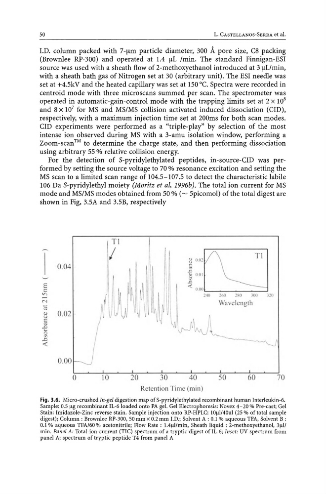
Fig. 3.6. Micro-crushed In-gel digestion map of S-pyridylethylated recombinant human Interleukin-6.
Sample: 0.5 I-tg recombinant IL-6loaded onto PA gel. Gel Electrophoresis: Novex 4-20 % Pre-cast; Gel
Stain: Imidazole-Zinc reverse stain. Sample injection onto RP-HPLC: IOI-tl!40ul (25% of total sample
digest); Column: Brownlee RP-300, 50 mm x 0.2 mm J.D.; Solvent A : 0.1 % aqueous TFA, Solvent B :
0.1 % aqueous TFA/60 % acetonitrile; Flow Rate: 1.41-tl!min, Sheath liquid: 2-methoxyethanol, 31-tl!
min. Panel A: Total-ion-current (TIC) spectrum of a tryptic digest of 11-6; Inset: UV spectrum from
panel A; spectrum of tryptic peptide T4 from panel A
L. CASTELLANOS-SERRA et al.
LD. column packed with 7-llm particle diameter, 300 A pore size, C8 packing
(Brownlee RP-300) and operated at 1.4 ilL Imin. The standard Finnigan-ESI
source was used with a sheath flow of 2-methoxyethanol introduced at 3 ilL/min,
with a sheath bath gas of Nitrogen set at 30 (arbitrary unit). The ESI needle was
set at +4.5kV and the heated capillary was set at 150°C. Spectra were recorded in
centroid mode with three microscans summed per scan. The spectrometer was
operated in automatic-gain-control mode with the trapping limits set at 2 x 10 8
and 8 x 10 7 for MS and MS/MS collision activated induced dissociation (Cm),
respectively, with a maximum injection time set at 200ms for both scan modes.
CID experiments were performed as a "triple-play" by selection of the most
intense ion observed during MS with a 3-amu isolation window, performing a
Zoom-scan ™ to determine the charge state, and then performing dissociation
using arbitrary 55 % relative collision energy.
For the detection of S-pyridylethylated peptides, in-source-Cm was performed by setting the source voltage to 70 % resonance excitation and setting the
MS scan to a limited scan range of 104.5-107.5 to detect the characteristic labile
106 Da S-pyridylethyl moiety (Moritz et aI, 1996b). The total ion current for MS
mode and MS/MS modes obtained from 50 % (~ 5picomol) of the total digest are
shown in Fig, 3.5A and 3.5B, respectively
T I
I
0.04
E
c
V)
N
~
<.)
0.02
u
c
'"
.0
....
0
(/)
.0
<:
0.00
0
10
20
~ 0.02
c
'"
.0
o 001
'"
.0
TI
< oOO L-_ _ _ _ _ _ _ ...J
240
!60
!KO
)011
120
Wavelength
30
40
50
60
Re tention Time (m in)
70
Fig. 3.6. Micro-crushed In-gel digestion map of S-pyridylethylated recombinant human Interleukin-6.
Sample: 0.5 I-tg recombinant IL-6loaded onto PA gel. Gel Electrophoresis: Novex 4-20 % Pre-cast; Gel
Stain: Imidazole-Zinc reverse stain. Sample injection onto RP-HPLC: IOI-tl!40ul (25% of total sample
digest); Column: Brownlee RP-300, 50 mm x 0.2 mm J.D.; Solvent A : 0.1 % aqueous TFA, Solvent B :
0.1 % aqueous TFA/60 % acetonitrile; Flow Rate: 1.41-tl!min, Sheath liquid: 2-methoxyethanol, 31-tl!
min. Panel A: Total-ion-current (TIC) spectrum of a tryptic digest of 11-6; Inset: UV spectrum from
panel A; spectrum of tryptic peptide T4 from panel A