

30
L. CASTELLANOS-SERRA et al.
This improved zinc staining has been termed Reverse-staining; the term has a
dual connotation, alluding both to the "reverse" character of the stain (background or negative stain) and to the fact that the process can be fully "reversed"
to the unstained -original- state. The procedure is being currently used by many
research groups for protein detection before MS analysis or in-gel digestion
(Cohen and Chait, 1997; Matsui et aI, 1997; Gevaert et aI, 1998; Scheler et aI, 1998)
and a staining kit is now commercially available. Recently, it was shown that
Reverse-staining is not limited to proteins, but this can be generally applied to
the sensitive visualization of other biomolecules - nucleic acids (Hardy et aI,
1996) and bacteriallipopolysaccharides (Hardy et aI, 1997) - on electrophoresis
gels.
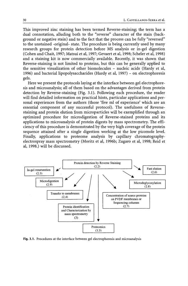
Here we present the protocols laying at the interface between gel electrophoresis and microanalysis; all of them based on the advantages derived from protein
detection by Reverse-staining (Fig. 3.1). Following each procedure, the reader
will find detailed information on practical hints, particular applications and personal experiences from the authors (those 'five ml of experience' which are an
essential component of any successful protocol). The usefulness of Reversestaining and protein elution from microparticles will be exemplified through an
optimized procedure for microdigestion of Reverse-stained proteins and its
applications to microanalysis of protein digests by mass spectrometry. The efficiency of this procedure is demonstrated by the very high coverage of the protein
sequence attained after a single digestion working at the low picomole level.
Finally, applications to proteome analysis by capillary chromatographyelectrospray mass spectrometry (Moritz et aI, 1996b; Zugaro et al, 1998; Reid et
aI, 1998.) will be discussed.
Protein detection by Reverse Staining
(2.2)
"'~----------------~
~
Transfer to membranes
(2.4)
Protein identification
and Characterization by
mass spectrometry
(3)
Proteomics
(3.3)
~
Microdeglycosylation
(2.8)
Concentration of scarce proteins
on PVDF membranes or
Sequencing columns
(2.7)
Fig. 3.1. Procedures at the interface between gel electrophoresis and microanalysis
L. CASTELLANOS-SERRA et al.
This improved zinc staining has been termed Reverse-staining; the term has a
dual connotation, alluding both to the "reverse" character of the stain (background or negative stain) and to the fact that the process can be fully "reversed"
to the unstained -original- state. The procedure is being currently used by many
research groups for protein detection before MS analysis or in-gel digestion
(Cohen and Chait, 1997; Matsui et aI, 1997; Gevaert et aI, 1998; Scheler et aI, 1998)
and a staining kit is now commercially available. Recently, it was shown that
Reverse-staining is not limited to proteins, but this can be generally applied to
the sensitive visualization of other biomolecules - nucleic acids (Hardy et aI,
1996) and bacteriallipopolysaccharides (Hardy et aI, 1997) - on electrophoresis
gels.
Here we present the protocols laying at the interface between gel electrophoresis and microanalysis; all of them based on the advantages derived from protein
detection by Reverse-staining (Fig. 3.1). Following each procedure, the reader
will find detailed information on practical hints, particular applications and personal experiences from the authors (those 'five ml of experience' which are an
essential component of any successful protocol). The usefulness of Reversestaining and protein elution from microparticles will be exemplified through an
optimized procedure for microdigestion of Reverse-stained proteins and its
applications to microanalysis of protein digests by mass spectrometry. The efficiency of this procedure is demonstrated by the very high coverage of the protein
sequence attained after a single digestion working at the low picomole level.
Finally, applications to proteome analysis by capillary chromatographyelectrospray mass spectrometry (Moritz et aI, 1996b; Zugaro et al, 1998; Reid et
aI, 1998.) will be discussed.
Protein detection by Reverse Staining
(2.2)
"'~----------------~
~
Transfer to membranes
(2.4)
Protein identification
and Characterization by
mass spectrometry
(3)
Proteomics
(3.3)
~
Microdeglycosylation
(2.8)
Concentration of scarce proteins
on PVDF membranes or
Sequencing columns
(2.7)
Fig. 3.1. Procedures at the interface between gel electrophoresis and microanalysis