

Proteoglycans: Biological Roles and Strategies
!
HA
Isolated PGs, Tissues, Cell cultures
1
Papain digestion
and/or alkaline borohydride
treatment
Separation between GAGs
I. Anion-exchange chromatography
-washing 0.1 M NaCi
-gradient elution: 0.1-1.2M
2. Gel-permeation chromatography
Characterization of isolated GAGs
i
GalAG. (CSIDS)
with chondroitinases +chandro-4- and
chondroitinases
ABC+AC
357
HS/heparin
Separate
digestions with
I
chondroitinases
ABC+AC
or hyalunonidase
streptomyces
HPLCorCE
Separate digestions j;;;:;:~inases
ABC, AC and B
-6-sulfatases
heparin
heparin Iyases /, II,
Iyases J.IJ.II1 III
• Determine HA from
Mi·nonSHA
HPLCorCE
• Determine total GalAG
content from6di-nonSCSIDS
Ion-pair HPLC or CE
• Identify HS or heparin
Ion-pair HPLC and CE
• Identify OS
• Determine sequence of OS
HPLCor E
• Determine disaccharide
composition (sulfation pattern)
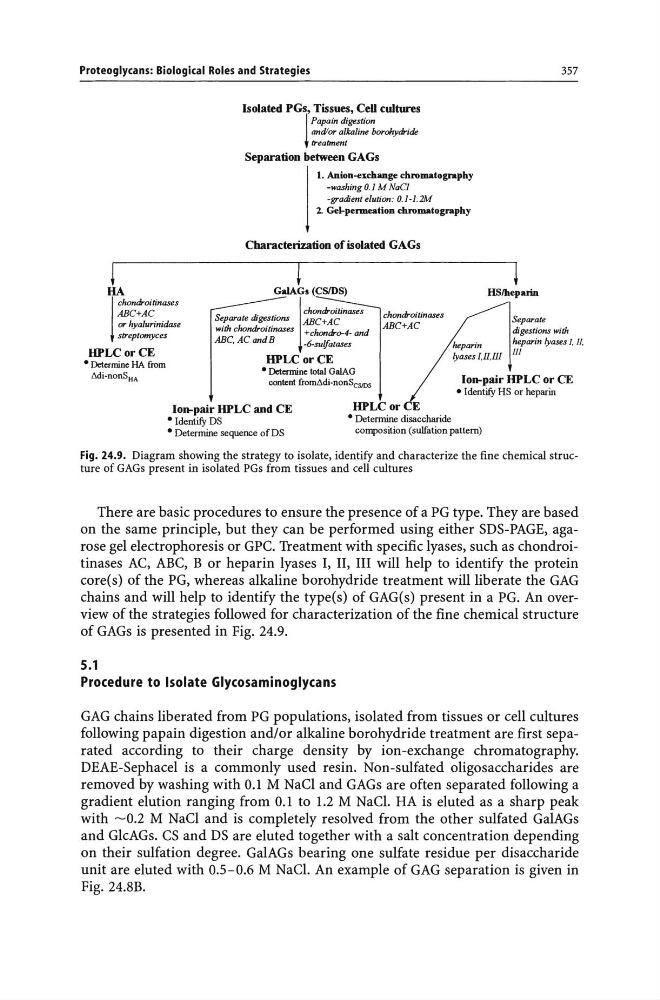
Fig. 24.9. Diagram showing the strategy to isolate, identify and characterize the fine chemical structure of GAGs present in isolated PGs from tissues and cell cultures
There are basic procedures to ensure the presence of a PG type. They are based
on the same principle, but they can be performed using either SDS-PAGE, agarose gel electrophoresis or GPC. Treatment with specific lyases, such as chondroitinases AC, ABC, B or heparin lyases I, II, III will help to identify the protein
core(s) of the PG, whereas alkaline borohydride treatment will liberate the GAG
chains and will help to identify the type(s) of GAG(s) present in a PG. An overview of the strategies followed for characterization of the fine chemical structure
of GAGs is presented in Fig. 24.9.
5.1
Procedure to Isolate Glycosaminoglycans
GAG chains liberated from PG populations, isolated from tissues or cell cultures
following papain digestion and/or alkaline borohydride treatment are first separated according to their charge density by ion-exchange chromatography.
DEAE-Sephacel is a commonly used resin. Non-sulfated oligo saccharides are
removed by washing with 0.1 M NaCI and GAGs are often separated following a
gradient elution ranging from 0.1 to 1.2 M NaCl. HA is eluted as a sharp peak
with -0.2 M NaCI and is completely resolved from the other sulfated GalAGs
and GlcAGs. CS and DS are eluted together with a salt concentration depending
on their sulfation degree. GalAGs bearing one sulfate residue per disaccharide
unit are eluted with 0.5-0.6 M NaCl. An example of GAG separation is given in
Fig. 24.BB.
!
HA
Isolated PGs, Tissues, Cell cultures
1
Papain digestion
and/or alkaline borohydride
treatment
Separation between GAGs
I. Anion-exchange chromatography
-washing 0.1 M NaCi
-gradient elution: 0.1-1.2M
2. Gel-permeation chromatography
Characterization of isolated GAGs
i
GalAG. (CSIDS)
with chondroitinases +chandro-4- and
chondroitinases
ABC+AC
357
HS/heparin
Separate
digestions with
I
chondroitinases
ABC+AC
or hyalunonidase
streptomyces
HPLCorCE
Separate digestions j;;;:;:~inases
ABC, AC and B
-6-sulfatases
heparin
heparin Iyases /, II,
Iyases J.IJ.II1 III
• Determine HA from
Mi·nonSHA
HPLCorCE
• Determine total GalAG
content from6di-nonSCSIDS
Ion-pair HPLC or CE
• Identify HS or heparin
Ion-pair HPLC and CE
• Identify OS
• Determine sequence of OS
HPLCor E
• Determine disaccharide
composition (sulfation pattern)
Fig. 24.9. Diagram showing the strategy to isolate, identify and characterize the fine chemical structure of GAGs present in isolated PGs from tissues and cell cultures
There are basic procedures to ensure the presence of a PG type. They are based
on the same principle, but they can be performed using either SDS-PAGE, agarose gel electrophoresis or GPC. Treatment with specific lyases, such as chondroitinases AC, ABC, B or heparin lyases I, II, III will help to identify the protein
core(s) of the PG, whereas alkaline borohydride treatment will liberate the GAG
chains and will help to identify the type(s) of GAG(s) present in a PG. An overview of the strategies followed for characterization of the fine chemical structure
of GAGs is presented in Fig. 24.9.
5.1
Procedure to Isolate Glycosaminoglycans
GAG chains liberated from PG populations, isolated from tissues or cell cultures
following papain digestion and/or alkaline borohydride treatment are first separated according to their charge density by ion-exchange chromatography.
DEAE-Sephacel is a commonly used resin. Non-sulfated oligo saccharides are
removed by washing with 0.1 M NaCI and GAGs are often separated following a
gradient elution ranging from 0.1 to 1.2 M NaCl. HA is eluted as a sharp peak
with -0.2 M NaCI and is completely resolved from the other sulfated GalAGs
and GlcAGs. CS and DS are eluted together with a salt concentration depending
on their sulfation degree. GalAGs bearing one sulfate residue per disaccharide
unit are eluted with 0.5-0.6 M NaCl. An example of GAG separation is given in
Fig. 24.BB.