

A Structural Model for the PS3 Complex with DNA Response Elements
263
3
~
~ 4
(b)
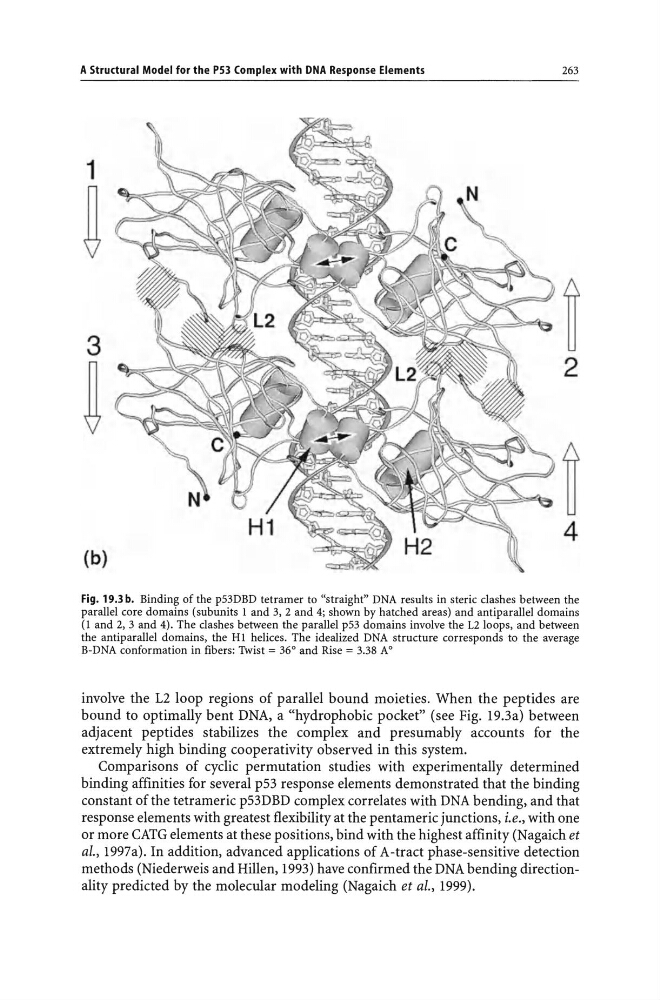
Fig. 19.3 b. Binding of the p53DBD tetramer to "straight" DNA results in steric clashes between the
parallel core domains (subunits I and 3, 2 and 4; shown by hatched areas) and antiparallel domains
(I and 2, 3 and 4). The clashes between the parallel p53 domains involve the L2 loops, and between
the antiparallel domains, the HI helices. The idealized DNA structure corresponds to the average
B-DNA conformation in fibers: Twist = 36° and Rise = 3.38 AO
involve the L2 loop regions of parallel bound moieties. When the peptides are
bound to optimally bent DNA, a "hydrophobic pocket" (see Fig. 19.3a) between
adjacent pep tides stabilizes the complex and presumably accounts for the
extremely high binding cooperativity observed in this system.
Comparisons of cyclic permutation studies with experimentally determined
binding affinities for several p53 response elements demonstrated that the binding
constant of the tetrameric p53DBD complex correlates with DNA bending, and that
response elements with greatest flexibility at the pentameric junctions, i.e., with one
or more CATG elements at these positions, bind with the highest affinity (Nagaich et
al., 1997a). In addition, advanced applications of A-tract phase-sensitive detection
methods (Niederweis and Hillen, 1993) have confirmed the DNA bending directionality predicted by the molecular modeling (Nagaich et al., 1999).
263
3
~
~ 4
(b)
Fig. 19.3 b. Binding of the p53DBD tetramer to "straight" DNA results in steric clashes between the
parallel core domains (subunits I and 3, 2 and 4; shown by hatched areas) and antiparallel domains
(I and 2, 3 and 4). The clashes between the parallel p53 domains involve the L2 loops, and between
the antiparallel domains, the HI helices. The idealized DNA structure corresponds to the average
B-DNA conformation in fibers: Twist = 36° and Rise = 3.38 AO
involve the L2 loop regions of parallel bound moieties. When the peptides are
bound to optimally bent DNA, a "hydrophobic pocket" (see Fig. 19.3a) between
adjacent pep tides stabilizes the complex and presumably accounts for the
extremely high binding cooperativity observed in this system.
Comparisons of cyclic permutation studies with experimentally determined
binding affinities for several p53 response elements demonstrated that the binding
constant of the tetrameric p53DBD complex correlates with DNA bending, and that
response elements with greatest flexibility at the pentameric junctions, i.e., with one
or more CATG elements at these positions, bind with the highest affinity (Nagaich et
al., 1997a). In addition, advanced applications of A-tract phase-sensitive detection
methods (Niederweis and Hillen, 1993) have confirmed the DNA bending directionality predicted by the molecular modeling (Nagaich et al., 1999).