

12
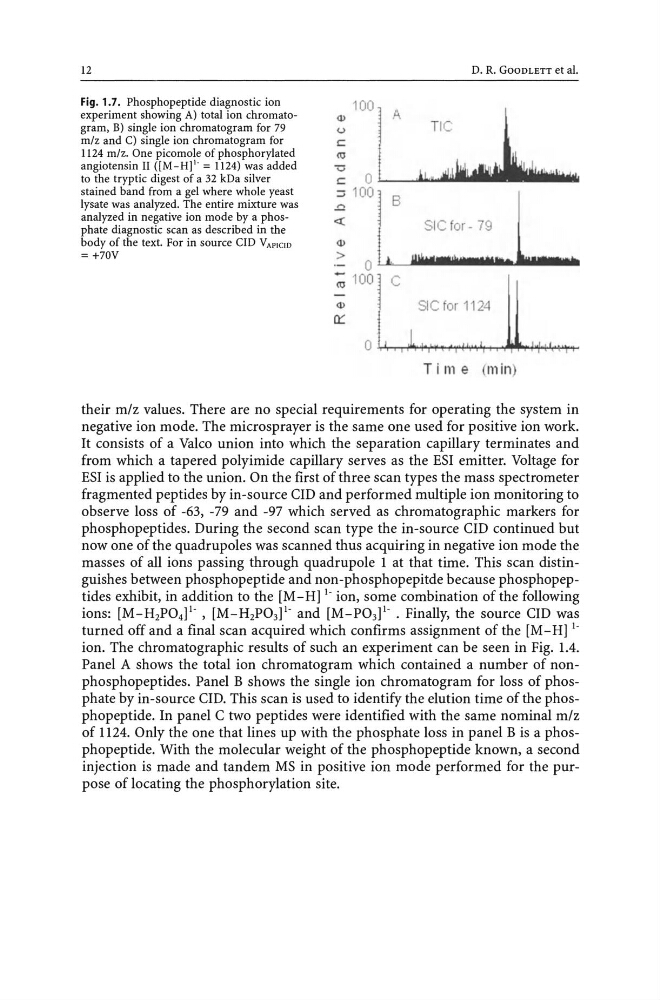
Fig. 1.7. Phosphopeptide diagnostic ion
experiment showing A) total ion chromatogram, B) single ion chromatogram for 79
mlz and C) single ion chromatogram for
1124 mlz. One picomole of phosphorylated
angiotensin II ([M_H]I. = 1124) was added
to the tryptic digest of a 32 kDa silver
stained band from a gel where whole yeast
lysate was analyzed. The entire mixture was
analyzed in negative ion mode by a phosphate diagnostic scan as described in the
body of the text. For in source CID VAPICID
= +70V
D. R. GOODLETT et aI.
_ Q ) : : ~ : : : 10
0
0 l L_ A----.JJ..i...lI _ _ _ l ... M
= _ , 1 '~:l ! ! I "'I! '+I + " ' ! : !"r l r ! ,
r: Ll J..> B~J & Y I l j ~ I I I I . : . f i l l l l : I I I I : .
- .. : I I ~ t ~ M ... ioJl .. ' lIs_lIInlilllb
('IJ 100 C
SIC for 1124
Time (min)
their m/z values. There are no special requirements for operating the system in
negative ion mode. The microsprayer is the same one used for positive ion work.
It consists of a Valco union into which the separation capillary terminates and
from which a tapered polyimide capillary serves as the ESI emitter. Voltage for
ESI is applied to the union. On the first of three scan types the mass spectrometer
fragmented peptides by in-source cm and performed multiple ion monitoring to
observe loss of -63, -79 and -97 which served as chromatographic markers for
phosphopeptides. During the second scan type the in-source cm continued but
now one of the quadrupoles was scanned thus acquiring in negative ion mode the
masses of all ions passing through quadrupole 1 at that time. This scan distinguishes between phosphopeptide and non-phosphopepitde because phosphopeptides exhibit, in addition to the [M - Hj!' ion, some combination of the following
ions: [M-H2P04P' , [M-H2P03j!' and [M-P03j!' . Finally, the source cm was
turned off and a final scan acquired which confirms assignment of the [M-Hj!'
ion. The chromatographic results of such an experiment can be seen in Fig. 1.4.
Panel A shows the total ion chromatogram which contained a number of nonphosphopeptides. Panel B shows the single ion chromatogram for loss of phosphate by in-source cm. This scan is used to identify the elution time of the phosphopeptide. In panel C two peptides were identified with the same nominal m/z
of 1124. Only the one that lines up with the phosphate loss in panel B is a phosphopeptide. With the molecular weight of the phosphopeptide known, a second
injection is made and tandem MS in positive ion mode performed for the purpose of locating the phosphorylation site.
Fig. 1.7. Phosphopeptide diagnostic ion
experiment showing A) total ion chromatogram, B) single ion chromatogram for 79
mlz and C) single ion chromatogram for
1124 mlz. One picomole of phosphorylated
angiotensin II ([M_H]I. = 1124) was added
to the tryptic digest of a 32 kDa silver
stained band from a gel where whole yeast
lysate was analyzed. The entire mixture was
analyzed in negative ion mode by a phosphate diagnostic scan as described in the
body of the text. For in source CID VAPICID
= +70V
D. R. GOODLETT et aI.
_ Q ) : : ~ : : : 10
0
0 l L_ A----.JJ..i...lI _ _ _ l ... M
= _ , 1 '~:l ! ! I "'I! '+I + " ' ! : !"r l r ! ,
r: Ll J..> B~J & Y I l j ~ I I I I . : . f i l l l l : I I I I : .
- .. : I I ~ t ~ M ... ioJl .. ' lIs_lIInlilllb
('IJ 100 C
SIC for 1124
Time (min)
their m/z values. There are no special requirements for operating the system in
negative ion mode. The microsprayer is the same one used for positive ion work.
It consists of a Valco union into which the separation capillary terminates and
from which a tapered polyimide capillary serves as the ESI emitter. Voltage for
ESI is applied to the union. On the first of three scan types the mass spectrometer
fragmented peptides by in-source cm and performed multiple ion monitoring to
observe loss of -63, -79 and -97 which served as chromatographic markers for
phosphopeptides. During the second scan type the in-source cm continued but
now one of the quadrupoles was scanned thus acquiring in negative ion mode the
masses of all ions passing through quadrupole 1 at that time. This scan distinguishes between phosphopeptide and non-phosphopepitde because phosphopeptides exhibit, in addition to the [M - Hj!' ion, some combination of the following
ions: [M-H2P04P' , [M-H2P03j!' and [M-P03j!' . Finally, the source cm was
turned off and a final scan acquired which confirms assignment of the [M-Hj!'
ion. The chromatographic results of such an experiment can be seen in Fig. 1.4.
Panel A shows the total ion chromatogram which contained a number of nonphosphopeptides. Panel B shows the single ion chromatogram for loss of phosphate by in-source cm. This scan is used to identify the elution time of the phosphopeptide. In panel C two peptides were identified with the same nominal m/z
of 1124. Only the one that lines up with the phosphate loss in panel B is a phosphopeptide. With the molecular weight of the phosphopeptide known, a second
injection is made and tandem MS in positive ion mode performed for the purpose of locating the phosphorylation site.