

A MALO 1-TOF Mass Spectrometry Approach to Investigate the Defense Reactions
171
Although a purification step introduces selectivity, the isolation of a specific product can be followed with the help of its molecular mass. The structural characterization can be performed using different techniques depending on the amount of
peptide available, the necessity to have a partial or a full sequence and the possibility to conduct molecular cloning experiments.
9
MALDI-TOF MS as a Tool to Investigate Inducible Tissue-Specific
Expression of the Drosomycin Gene
We have also investigated the possible expression of Drosophila antimicrobial
peptides in barrier epithelia using the antifungal peptide drosomycin as a model.
Using a transgenic reporter system based on the green fluorescent protein (GFP
from the jellyfish Aequorea victori, Chalfie et al. 1994), it has been shown that the
reporter gene drosomycin-GFP was expressed in different epithelial tissues, for
example in tracheal trunks of some experimentally uninfected Drosophila larvae
(for more details, see Ferrandon et al. 1998). However, to be certain that the GFP
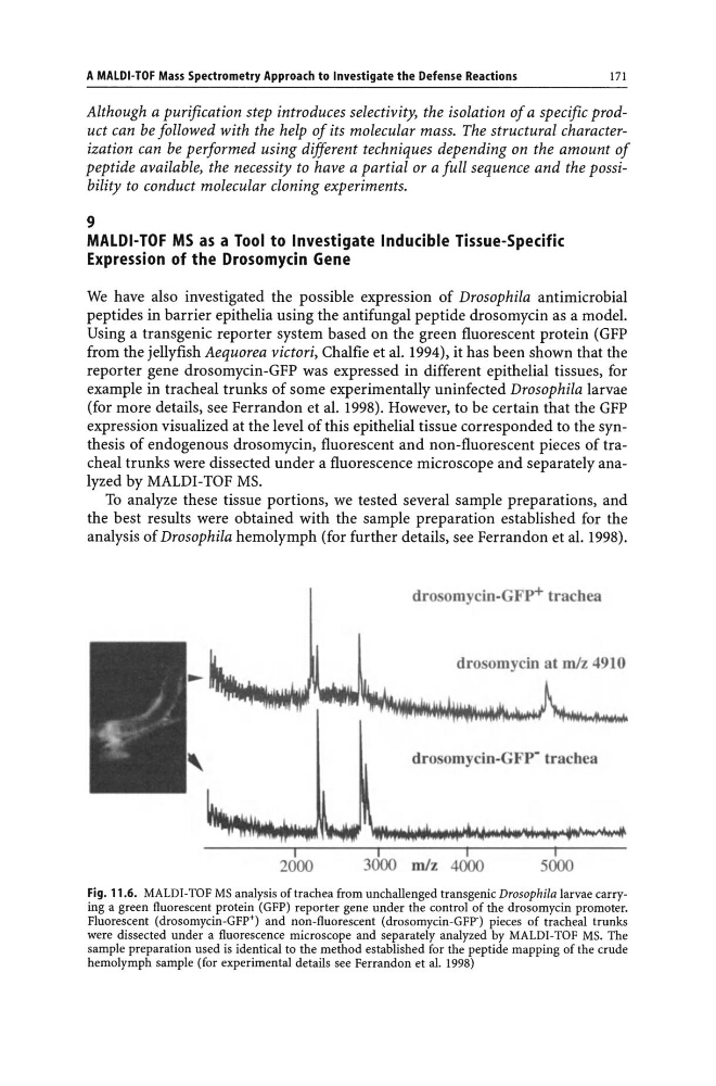
expression visualized at the level of this epithelial tissue corresponded to the synthesis of endogenous drosomycin, fluorescent and non-fluorescent pieces of tracheal trunks were dissected under a fluorescence microscope and separately analyzed by MALDI-TOF MS.
To analyze these tissue portions, we tested several sample preparations, and
the best results were obtained with the sample preparation established for the
analysis of Drosophila hemolymph (for further details, see Ferrandon et al. 1998).
drosomycin-GFP+ trachea
drosomycin at mJz 4910
dro omycin-GFP· trachea
I
I
I
3000 mJz 4000
5000
Fig. 11.6. MALDI-TOF MS analysis of trachea from unchallenged transgenic Drosophila larvae carrying a green fluorescent protein (GFP) reporter gene under the control of the drosomycin promoter.
Fluorescent (drosomycin-GFP+) and non-fluorescent (drosomycin-GFP-) pieces of tracheal trunks
were dissected under a fluorescence microscope and separately analyzed by MALDI-TOF MS. The
sample preparation used is identical to the method established for the peptide mapping of the crude
hemolymph sample (for experimental details see Ferrandon et al. 1998)
171
Although a purification step introduces selectivity, the isolation of a specific product can be followed with the help of its molecular mass. The structural characterization can be performed using different techniques depending on the amount of
peptide available, the necessity to have a partial or a full sequence and the possibility to conduct molecular cloning experiments.
9
MALDI-TOF MS as a Tool to Investigate Inducible Tissue-Specific
Expression of the Drosomycin Gene
We have also investigated the possible expression of Drosophila antimicrobial
peptides in barrier epithelia using the antifungal peptide drosomycin as a model.
Using a transgenic reporter system based on the green fluorescent protein (GFP
from the jellyfish Aequorea victori, Chalfie et al. 1994), it has been shown that the
reporter gene drosomycin-GFP was expressed in different epithelial tissues, for
example in tracheal trunks of some experimentally uninfected Drosophila larvae
(for more details, see Ferrandon et al. 1998). However, to be certain that the GFP
expression visualized at the level of this epithelial tissue corresponded to the synthesis of endogenous drosomycin, fluorescent and non-fluorescent pieces of tracheal trunks were dissected under a fluorescence microscope and separately analyzed by MALDI-TOF MS.
To analyze these tissue portions, we tested several sample preparations, and
the best results were obtained with the sample preparation established for the
analysis of Drosophila hemolymph (for further details, see Ferrandon et al. 1998).
drosomycin-GFP+ trachea
drosomycin at mJz 4910
dro omycin-GFP· trachea
I
I
I
3000 mJz 4000
5000
Fig. 11.6. MALDI-TOF MS analysis of trachea from unchallenged transgenic Drosophila larvae carrying a green fluorescent protein (GFP) reporter gene under the control of the drosomycin promoter.
Fluorescent (drosomycin-GFP+) and non-fluorescent (drosomycin-GFP-) pieces of tracheal trunks
were dissected under a fluorescence microscope and separately analyzed by MALDI-TOF MS. The
sample preparation used is identical to the method established for the peptide mapping of the crude
hemolymph sample (for experimental details see Ferrandon et al. 1998)