

140
E. LEHMANN and R. ZENOBI
(Gruic-Sovulj et al. 1997; Lehmann 1996). For this purpose, control experiments
had to be designed. Furthermore, we wanted to establish a correlation between
the complexation behavior in solution and in the MALDI spectra. A detailed
report of the experiments described in this chapter can be found in two recent
papers (Lehmann and Zenobi 1998, Lehmann et al. 1999)
2
Results and Discussion
2.1
Zinc Finger Peptide Complexation with Metal Ions
We investigated an 18-residue zinc finger peptide of CCHC type (CCHC = CysXz-Cys-X4-His-~-Cys, X = variable amino acid) with the sequence acVKCFNCGKEGHIARNCRA-OH corresponding to the first zinc finger domain from the
gag protein p55 of human immunodeficiency virus type 1 (HIV-l), called p55Fl.
The apo-peptide has no defined secondary structure. Metal ion binding induces
peptide folding which is required for the interaction with nucleic acids (Lam et
al. 1994). CCHC type peptides bind Zn 2 + very tightly. In the resulting complex,
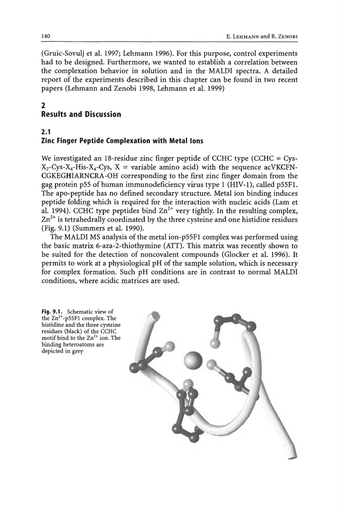
Zn 2 + is tetrahedrally coordinated by the three cysteine and one histidine residues
(Fig. 9.1) (Summers et al. 1990).
The MALD! MS analysis of the metal ion-p55Fl complex was performed using
the basic matrix 6-aza-2-thiothymine (ATT). This matrix was recently shown to
be suited for the detection of non covalent compounds (Glocker et al. 1996). It
permits to work at a physiological pH of the sample solution, which is necessary
for complex formation. Such pH conditions are in contrast to normal MALDI
conditions, where acidic matrices are used.
Fig. 9.1. Schematic view of
the Zn 2 + -p55F1 complex. The
histidine and the three cysteine
residues (black) of the CCRC
motif bind to the Zn2+ ion. The
binding heteroatoms are
depicted in grey
E. LEHMANN and R. ZENOBI
(Gruic-Sovulj et al. 1997; Lehmann 1996). For this purpose, control experiments
had to be designed. Furthermore, we wanted to establish a correlation between
the complexation behavior in solution and in the MALDI spectra. A detailed
report of the experiments described in this chapter can be found in two recent
papers (Lehmann and Zenobi 1998, Lehmann et al. 1999)
2
Results and Discussion
2.1
Zinc Finger Peptide Complexation with Metal Ions
We investigated an 18-residue zinc finger peptide of CCHC type (CCHC = CysXz-Cys-X4-His-~-Cys, X = variable amino acid) with the sequence acVKCFNCGKEGHIARNCRA-OH corresponding to the first zinc finger domain from the
gag protein p55 of human immunodeficiency virus type 1 (HIV-l), called p55Fl.
The apo-peptide has no defined secondary structure. Metal ion binding induces
peptide folding which is required for the interaction with nucleic acids (Lam et
al. 1994). CCHC type peptides bind Zn 2 + very tightly. In the resulting complex,
Zn 2 + is tetrahedrally coordinated by the three cysteine and one histidine residues
(Fig. 9.1) (Summers et al. 1990).
The MALD! MS analysis of the metal ion-p55Fl complex was performed using
the basic matrix 6-aza-2-thiothymine (ATT). This matrix was recently shown to
be suited for the detection of non covalent compounds (Glocker et al. 1996). It
permits to work at a physiological pH of the sample solution, which is necessary
for complex formation. Such pH conditions are in contrast to normal MALDI
conditions, where acidic matrices are used.
Fig. 9.1. Schematic view of
the Zn 2 + -p55F1 complex. The
histidine and the three cysteine
residues (black) of the CCRC
motif bind to the Zn2+ ion. The
binding heteroatoms are
depicted in grey