

Elucidation of Functionally Significant Structural Modifications
129
Table 8.2 (Continued)
Fragment
m/z
Predicted
Observed
Difference
Observed
Difference
V466-84
VRI82-6409
V466-84
66-84
y7
590.6
663.0
72.4
y8
691.7
764.0
72.3
691.4
-0.3
y9
792.8
865.3
72.5
792.3
-0.5
z9
775.8
775.1
-0.7
ylO
892.0
965.0
73
yll
979.1
1051.4
72.3
z14
1332.4
1405.4
73
y15
1505.6
1577.6
72
1505.2
-0.4
y16
1661.8
1732.7
70.9
1660.1
-1.7
y17
1775.0
1846.4
71.4
y18
1862.1
1934.4
72.3
1. These ions are diagnostic of a C-terminal glutamic acid and C-terminal glycine for the VRI 82-6409
and V 4 sequences, respectively.
2. These data were diagnostic of a difference of a glycine and glutamic acid within the last 3 residues
at the C-termini of the V 4 and VRI 82-6409 sequences.
a.i.
6500
6000
5500
5000
4500
4000
3500
3000
2500
2000
1500
1000
500
o
QNKPPSKPNNDFHFEVFNFVPCSICSNNPTCWAICK
I
I
I
I
I
I
I
I
L ______ 1 _______________ l. __________ I
4108.1
4125.4
3600
3800
m/z
4000
4200
4400
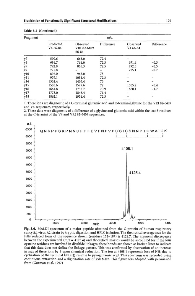
Fig. 8.6. MALD! spectrum of a major peptide obtained from the G-protein of human respiratory
syncytial virus A2 strain by tryptic digestion and HPLC isolation. The theoretical average m/z for the
fully reduced form of the sequence shown (residues 152-187) is 4128.7. The apparent discrepancy
between the experimental (m/z = 4125.4) and theoretical masses would be accounted for if the four
cysteine residues are involved in disulfide linkages, these bonds are shown as broken lines to indicate
that this data does not define the linkage pattern. This was confirmed by observation of an increase
in m/z of these ions by 4 upon chemical reduction. The ion at 4108.1 represents loss of NH, due to
cyclization of the terminal Gin (Q) residue to pyroglutamic acid. This spectrum was recorded using
continuous extraction and a digitization rate of 250 MHz. This figure was adapted with permission
from (Gorman et al. 1997)
129
Table 8.2 (Continued)
Fragment
m/z
Predicted
Observed
Difference
Observed
Difference
V466-84
VRI82-6409
V466-84
66-84
y7
590.6
663.0
72.4
y8
691.7
764.0
72.3
691.4
-0.3
y9
792.8
865.3
72.5
792.3
-0.5
z9
775.8
775.1
-0.7
ylO
892.0
965.0
73
yll
979.1
1051.4
72.3
z14
1332.4
1405.4
73
y15
1505.6
1577.6
72
1505.2
-0.4
y16
1661.8
1732.7
70.9
1660.1
-1.7
y17
1775.0
1846.4
71.4
y18
1862.1
1934.4
72.3
1. These ions are diagnostic of a C-terminal glutamic acid and C-terminal glycine for the VRI 82-6409
and V 4 sequences, respectively.
2. These data were diagnostic of a difference of a glycine and glutamic acid within the last 3 residues
at the C-termini of the V 4 and VRI 82-6409 sequences.
a.i.
6500
6000
5500
5000
4500
4000
3500
3000
2500
2000
1500
1000
500
o
QNKPPSKPNNDFHFEVFNFVPCSICSNNPTCWAICK
I
I
I
I
I
I
I
I
L ______ 1 _______________ l. __________ I
4108.1
4125.4
3600
3800
m/z
4000
4200
4400
Fig. 8.6. MALD! spectrum of a major peptide obtained from the G-protein of human respiratory
syncytial virus A2 strain by tryptic digestion and HPLC isolation. The theoretical average m/z for the
fully reduced form of the sequence shown (residues 152-187) is 4128.7. The apparent discrepancy
between the experimental (m/z = 4125.4) and theoretical masses would be accounted for if the four
cysteine residues are involved in disulfide linkages, these bonds are shown as broken lines to indicate
that this data does not define the linkage pattern. This was confirmed by observation of an increase
in m/z of these ions by 4 upon chemical reduction. The ion at 4108.1 represents loss of NH, due to
cyclization of the terminal Gin (Q) residue to pyroglutamic acid. This spectrum was recorded using
continuous extraction and a digitization rate of 250 MHz. This figure was adapted with permission
from (Gorman et al. 1997)