

112
B. BERSCH et al.
- •
- -1.0
0.8
C\J
0.6
(j)
0.4
0.2
30
Vi
.e: 20
p
10
10
N
8
;S
6
x
(1)
4
a::
2
0
20
40
60
80
100
120
residue number
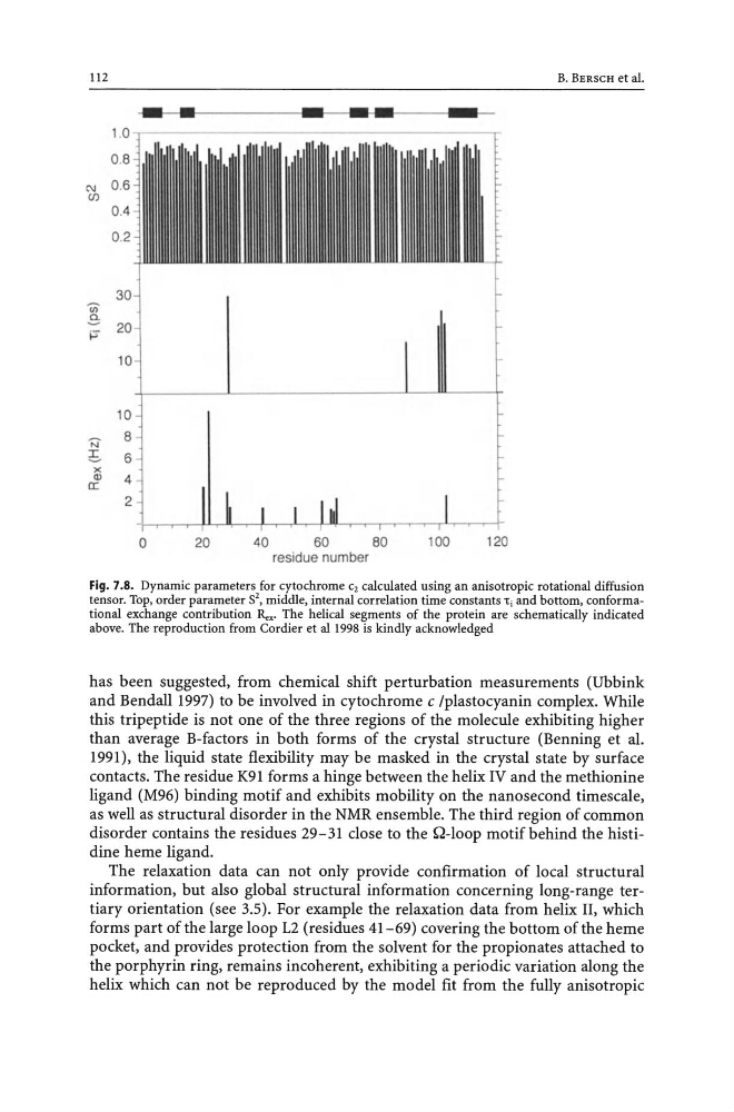
Fig. 7.S. Dynamic parameters for cytochrome C2 calculated using an anisotropic rotational diffusion
tensor. Top, order parameter 52, middle, internal correlation time constants 1:, and bottom, conformational exchange contribution Rex. The helical segments of the protein are schematically indicated
above. The reproduction from Cordier et al 1998 is kindly acknowledged
has been suggested, from chemical shift perturbation measurements (Ubbink
and Bendall 1997) to be involved in cytochrome c Iplastocyanin complex. While
this tripeptide is not one of the three regions of the molecule exhibiting higher
than average B-factors in both forms of the crystal structure (Benning et al.
1991), the liquid state flexibility may be masked in the crystal state by surface
contacts. The residue K91 forms a hinge between the helix IV and the methionine
ligand (M96) binding motif and exhibits mobility on the nanosecond timescale,
as well as structural disorder in the NMR ensemble. The third region of common
disorder contains the residues 29-31 close to the Q-Ioop motif behind the histidine heme ligand.
The relaxation data can not only provide confirmation of local structural
information, but also global structural information concerning long-range tertiary orientation (see 3.5). For example the relaxation data from helix II, which
forms part of the large loop L2 (residues 41-69) covering the bottom of the heme
pocket, and provides protection from the solvent for the propionates attached to
the porphyrin ring, remains incoherent, exhibiting a periodic variation along the
helix which can not be reproduced by the model fit from the fully anisotropic
B. BERSCH et al.
- •
- -1.0
0.8
C\J
0.6
(j)
0.4
0.2
30
Vi
.e: 20
p
10
10
N
8
;S
6
x
(1)
4
a::
2
0
20
40
60
80
100
120
residue number
Fig. 7.S. Dynamic parameters for cytochrome C2 calculated using an anisotropic rotational diffusion
tensor. Top, order parameter 52, middle, internal correlation time constants 1:, and bottom, conformational exchange contribution Rex. The helical segments of the protein are schematically indicated
above. The reproduction from Cordier et al 1998 is kindly acknowledged
has been suggested, from chemical shift perturbation measurements (Ubbink
and Bendall 1997) to be involved in cytochrome c Iplastocyanin complex. While
this tripeptide is not one of the three regions of the molecule exhibiting higher
than average B-factors in both forms of the crystal structure (Benning et al.
1991), the liquid state flexibility may be masked in the crystal state by surface
contacts. The residue K91 forms a hinge between the helix IV and the methionine
ligand (M96) binding motif and exhibits mobility on the nanosecond timescale,
as well as structural disorder in the NMR ensemble. The third region of common
disorder contains the residues 29-31 close to the Q-Ioop motif behind the histidine heme ligand.
The relaxation data can not only provide confirmation of local structural
information, but also global structural information concerning long-range tertiary orientation (see 3.5). For example the relaxation data from helix II, which
forms part of the large loop L2 (residues 41-69) covering the bottom of the heme
pocket, and provides protection from the solvent for the propionates attached to
the porphyrin ring, remains incoherent, exhibiting a periodic variation along the
helix which can not be reproduced by the model fit from the fully anisotropic