

110
B. BERSCH et aJ.
1998). Cytochrome C2 has a broad distribution of helical motifs in angular space
permitting the precise determination of the anisotropic diffusion tensor, and
thus an accurate parameterization of the local motions of the peptide chain. The
precision of the analysis of the anisotropic diffusion tensor has been estimated
using Monte-Carlo sampling methods, made possible by an efficient simulated
annealing algorithm developed in our laboratory.
4.2.1
Characterization of Local-Motion and Overall Tumbling
Use of a spectral density function assuming an isotropic rotational diffusion tensor in the Lipari-Szabo modelfree approach reveals a highly compact protein.
However certain irregularities indicate that this model is imperfect - nearly
thirty residues require a more complex model than the most rigid one, 10 residues require a second motion on an intermediary timescale and 12 a chemical
exchange term. The situation of many of these residues in regions of secondary
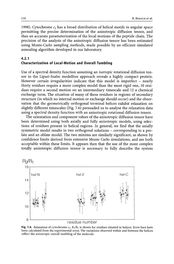
structure (in which no internal motion or exchange should occur) and the observation that the geometrically orthogonal terminal helices exhibit relaxation on
slightly different timescales (Fig. 7.6) persuaded us to analyse the relaxation data
using a spectral density function with an anisotropic rotational diffusion tensor.
The orientation and component values of the anisotropic diffusion tensor have
been determined using both axially and fully anisotropic models, using selections of residues present in helical regions. In general, we find that the axially
symmetric model results in two orthogonal solutions - corresponding to a prolate and an oblate model. The two minima are similarly significant, as shown by
confidence limits derived from extensive Monte Carlo simulations, and are both
acceptable within these limits. It appears then that the use of the more complex
totally anisotropic diffusion tensor is necessary to fully describe the system
RtR1
16.------------------------------------------,
hel-N
hel-2
hel-C
14
12
,L
10 ~,
~.
8L---------------~~----~--------------~
residue number
Fig. 7.6. Relaxation of cytochrome c,. R,/R J is shown for residues situated in helices. Error bars have
been calculated from the experimental error. The variations observed within and between the helices
reflect the anisotropic overall tumbling of the molecule
B. BERSCH et aJ.
1998). Cytochrome C2 has a broad distribution of helical motifs in angular space
permitting the precise determination of the anisotropic diffusion tensor, and
thus an accurate parameterization of the local motions of the peptide chain. The
precision of the analysis of the anisotropic diffusion tensor has been estimated
using Monte-Carlo sampling methods, made possible by an efficient simulated
annealing algorithm developed in our laboratory.
4.2.1
Characterization of Local-Motion and Overall Tumbling
Use of a spectral density function assuming an isotropic rotational diffusion tensor in the Lipari-Szabo modelfree approach reveals a highly compact protein.
However certain irregularities indicate that this model is imperfect - nearly
thirty residues require a more complex model than the most rigid one, 10 residues require a second motion on an intermediary timescale and 12 a chemical
exchange term. The situation of many of these residues in regions of secondary
structure (in which no internal motion or exchange should occur) and the observation that the geometrically orthogonal terminal helices exhibit relaxation on
slightly different timescales (Fig. 7.6) persuaded us to analyse the relaxation data
using a spectral density function with an anisotropic rotational diffusion tensor.
The orientation and component values of the anisotropic diffusion tensor have
been determined using both axially and fully anisotropic models, using selections of residues present in helical regions. In general, we find that the axially
symmetric model results in two orthogonal solutions - corresponding to a prolate and an oblate model. The two minima are similarly significant, as shown by
confidence limits derived from extensive Monte Carlo simulations, and are both
acceptable within these limits. It appears then that the use of the more complex
totally anisotropic diffusion tensor is necessary to fully describe the system
RtR1
16.------------------------------------------,
hel-N
hel-2
hel-C
14
12
,L
10 ~,
~.
8L---------------~~----~--------------~
residue number
Fig. 7.6. Relaxation of cytochrome c,. R,/R J is shown for residues situated in helices. Error bars have
been calculated from the experimental error. The variations observed within and between the helices
reflect the anisotropic overall tumbling of the molecule