

50
TAKEHlKO KO]l
taining requires optimization of the protocol depending upon
the stability of an epitope and the specimens which were not processed appropriately for each antigen or antibody to be investigated could result in false-negative or underestimated findings
(Ruizeveld de Winter et al. 1991; Slayden et al. 1995; Ehara et al.
1996). Practically, however, the optimal conditions cannot always be guaranteed for immunostaining with an antibody.
Thus, the use of in situ hybridization for mRNA has been indispensable not only for assessment of the expression of specific
mRNA, but also for verification of in situ synthesis of specific
proteins (Koji and Nakane 1996).
In situ hybridization depends upon base-pairing between target nucleic acids in cells or tissues and probe nucleic acids and its
conditions have been already optimized. The probe must be labeled with either radioactive or nonradioactive molecules to reveal the sites of hybridization. Originally, the probe nucleic acids
were labeled with radioisotopes such as 3H, 35S, 32p, 33p and 125 1
and the sites of hybrids were visualized by autoradiography. As
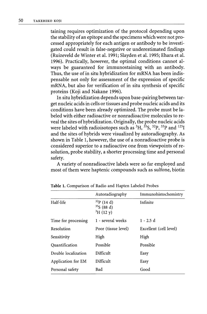
shown in Table 1, however, the use of a nonradioactive probe is
considered superior to a radioactive one from viewpoints of resolution, probe stability, a shorter processing time and personal
safety.
A variety of nonradioactive labels were so far employed and
most of them were haptenic compounds such as sulfone, biotin
Table 1. Comparison of Radio and Hapten Labeled Probes
Autoradiography
Half-life
32p (14 d)
35S (88 d)
3H (12 y)
Time for processing
1 - several weeks
Resolution
Poor (tissue level)
Sensitivity
High
Quantification
Possible
Double localization
Difficult
Application for EM
Difficult
Personal safety
Bad
Immunohistochemistry
Infinite
1 - 2.5 d
Excellent (cell level)
High
Possible
Easy
Easy
Good
TAKEHlKO KO]l
taining requires optimization of the protocol depending upon
the stability of an epitope and the specimens which were not processed appropriately for each antigen or antibody to be investigated could result in false-negative or underestimated findings
(Ruizeveld de Winter et al. 1991; Slayden et al. 1995; Ehara et al.
1996). Practically, however, the optimal conditions cannot always be guaranteed for immunostaining with an antibody.
Thus, the use of in situ hybridization for mRNA has been indispensable not only for assessment of the expression of specific
mRNA, but also for verification of in situ synthesis of specific
proteins (Koji and Nakane 1996).
In situ hybridization depends upon base-pairing between target nucleic acids in cells or tissues and probe nucleic acids and its
conditions have been already optimized. The probe must be labeled with either radioactive or nonradioactive molecules to reveal the sites of hybridization. Originally, the probe nucleic acids
were labeled with radioisotopes such as 3H, 35S, 32p, 33p and 125 1
and the sites of hybrids were visualized by autoradiography. As
shown in Table 1, however, the use of a nonradioactive probe is
considered superior to a radioactive one from viewpoints of resolution, probe stability, a shorter processing time and personal
safety.
A variety of nonradioactive labels were so far employed and
most of them were haptenic compounds such as sulfone, biotin
Table 1. Comparison of Radio and Hapten Labeled Probes
Autoradiography
Half-life
32p (14 d)
35S (88 d)
3H (12 y)
Time for processing
1 - several weeks
Resolution
Poor (tissue level)
Sensitivity
High
Quantification
Possible
Double localization
Difficult
Application for EM
Difficult
Personal safety
Bad
Immunohistochemistry
Infinite
1 - 2.5 d
Excellent (cell level)
High
Possible
Easy
Easy
Good