

44
YOSHINORI OTSUKI AND YUKO ITO
600.00
P< 0.01
P<0.05
500.00
!!:
P
400.00
-- ...
.8
e ::s
5
300.00
~
'1il
,g 200.00
t>I)
c
:.=
~ 100.00
~
0.00
......
.'tj
4
<.i
i
....
:::::
0'
0'
~
8
tS;
tS;
~
2
15
~
J:J
~
oS
it
~
.:y
s
f2
0
t
:;£
~
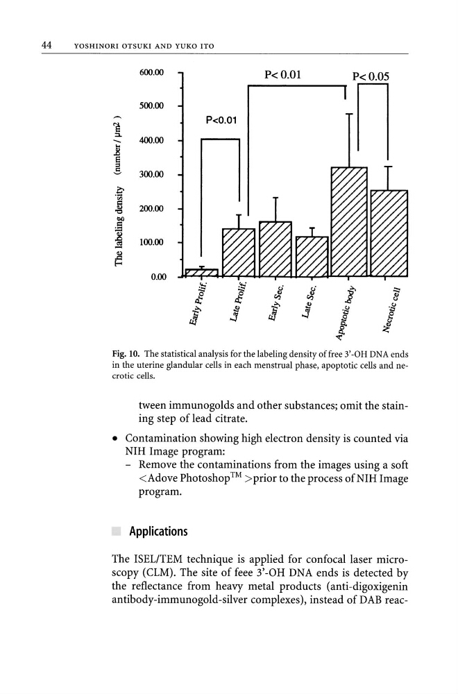
Fig. 10. The statistical analysis for the labeling density of free 3' -OH DNA ends
in the uterine glandular cells in each menstrual phase, apoptotic cells and necrotic cells.
tween immunogolds and other substances; omit the staining step of lead citrate.
• Contamination showing high electron density is counted via
NIH Image program:
- Remove the contaminations from the images using a soft
prior to the process of NIH Image
program.
Applications
The ISELITEM technique is applied for confocal laser microscopy (CLM). The site of feee 3'-OH DNA ends is detected by
the reflectance from heavy metal products (anti-digoxigenin
antibody-immunogold-silver complexes), instead of DAB reac-
YOSHINORI OTSUKI AND YUKO ITO
600.00
P< 0.01
P<0.05
500.00
!!:
P
400.00
-- ...
.8
e ::s
5
300.00
~
'1il
,g 200.00
t>I)
c
:.=
~ 100.00
~
0.00
......
.'tj
4
<.i
i
....
:::::
0'
0'
~
8
tS;
tS;
~
2
15
~
J:J
~
oS
it
~
.:y
s
f2
0
t
:;£
~
Fig. 10. The statistical analysis for the labeling density of free 3' -OH DNA ends
in the uterine glandular cells in each menstrual phase, apoptotic cells and necrotic cells.
tween immunogolds and other substances; omit the staining step of lead citrate.
• Contamination showing high electron density is counted via
NIH Image program:
- Remove the contaminations from the images using a soft
program.
Applications
The ISELITEM technique is applied for confocal laser microscopy (CLM). The site of feee 3'-OH DNA ends is detected by
the reflectance from heavy metal products (anti-digoxigenin
antibody-immunogold-silver complexes), instead of DAB reac-