

240
TAKEHIKO KOT!
Development of Southwestern histochemistry
During our efforts to optimize the protocol for nonradioactive in
situ hybridization using rat pituitary sections in 1985, we encountered a strange phenomenon. In an experiment, we hybridized the pituitary sections with dinitrophenyl (DNP)-labeled
proopiomelanocortin (POMC) cDNA and the expression of
POMC mRNA was analyzed enzyme-immunohistochemically
with anti-DNP antibody. As shown in Fig. 2, the specific staining
ofPOMC mRNA was found in intermediate pituitary cells as well
as some cells of anterior pituitary at a relatively high concentration of proteinase K. Surprisingly, a bacterial plasmid pBR 322
DNA, which was used as a negative control probe, gave rise to
some staining in a very similar distribution pattern to that of
POMC mRNA, but at low concentrations of proteinase K. Later,
we realized that the distribution of the "specific" protease- sensitive staining with pBR 322 DNA was quite similar to that of
glucocorticoid receptor (GR). Therefore, it was really exciting
ds o11go-r:tiA
~
": .
...
+
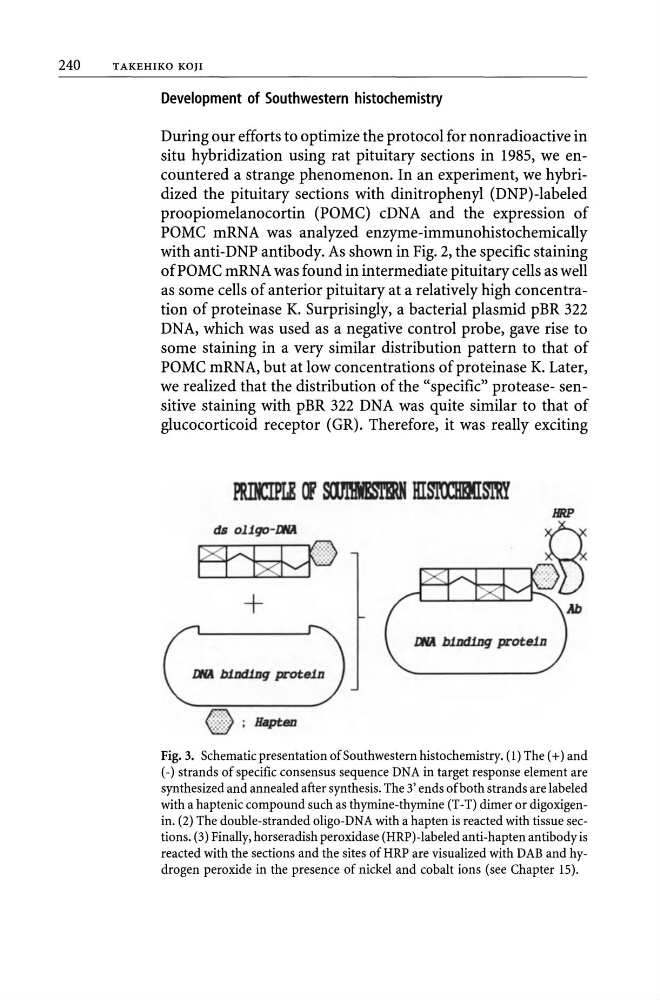
Fig. 3. Schematic presentation of Southwestern histochemistry. (1) The (+) and
(-) strands of specific consensus sequence DNA in target response element are
synthesized and annealed after synthesis. The 3' ends of both strands are labeled
with a haptenic compound such as thymine-thymine (T -T) dimer or digoxigenin. (2) The double-stranded oligo-DNA with a hapten is reacted with tissue sections. (3) Finally, horseradish peroxidase (HRP)-labeled anti-hapten antibody is
reacted with the sections and the sites ofHRP are visualized with DAB and hydrogen peroxide in the presence of nickel and cobalt ions (see Chapter 15).
TAKEHIKO KOT!
Development of Southwestern histochemistry
During our efforts to optimize the protocol for nonradioactive in
situ hybridization using rat pituitary sections in 1985, we encountered a strange phenomenon. In an experiment, we hybridized the pituitary sections with dinitrophenyl (DNP)-labeled
proopiomelanocortin (POMC) cDNA and the expression of
POMC mRNA was analyzed enzyme-immunohistochemically
with anti-DNP antibody. As shown in Fig. 2, the specific staining
ofPOMC mRNA was found in intermediate pituitary cells as well
as some cells of anterior pituitary at a relatively high concentration of proteinase K. Surprisingly, a bacterial plasmid pBR 322
DNA, which was used as a negative control probe, gave rise to
some staining in a very similar distribution pattern to that of
POMC mRNA, but at low concentrations of proteinase K. Later,
we realized that the distribution of the "specific" protease- sensitive staining with pBR 322 DNA was quite similar to that of
glucocorticoid receptor (GR). Therefore, it was really exciting
ds o11go-r:tiA
~
": .
...
+
Fig. 3. Schematic presentation of Southwestern histochemistry. (1) The (+) and
(-) strands of specific consensus sequence DNA in target response element are
synthesized and annealed after synthesis. The 3' ends of both strands are labeled
with a haptenic compound such as thymine-thymine (T -T) dimer or digoxigenin. (2) The double-stranded oligo-DNA with a hapten is reacted with tissue sections. (3) Finally, horseradish peroxidase (HRP)-labeled anti-hapten antibody is
reacted with the sections and the sites ofHRP are visualized with DAB and hydrogen peroxide in the presence of nickel and cobalt ions (see Chapter 15).